I. What are the Cardiovascular and Lymphatic Systems
Think of the cardiovascular and lymphatic systems as the body’s transportation and defense networks. They work in tandem to circulate blood and lymph throughout the body ensuring that nutrients, oxygen, and immune cells reach every part of your body!
While the cardiovascular system is more widely known due to its role in hearth health and circulation, the lymphatic system plays an equally crucial role in maintaining immune function and fluid balance. In this article, we’ll cover some of the main structures associated with the cardiovascular and lymphatic system as well as accompanying functions!
II. Structural Components of the Cardiovascular and Lymphatic System
We’ll try to keep our covering of the different structural components of the cardiovascular and lymphatic system to the main components without going too deep in detail in regards to the more specific histology and anatomy of these structures.
Rather, we’ll keep our focus and discussion to the major, high yield breakdown of the systems with a bigger emphasis on their physiological applications!
A. Major Structures of the Cardiovascular System
The cardiovascular system contains 3 main, major components: the heart, blood vessels, and blood. The heart generates the pumping force to allow for the flow of blood throughout the body via the blood vessels.
I. Heart
A majority of our discussion will revolve around the heart due to its central importance of generating the pressure gradient for blood flow as well as its anatomical and physiological complexity.
Located towards the left side of the chest, the heart is a muscular organ which contains 4 chambers with distinct separations from one another. The right and left atria (sing. atrium) are the top chambers while the right and left ventricles are the bottom chambers.
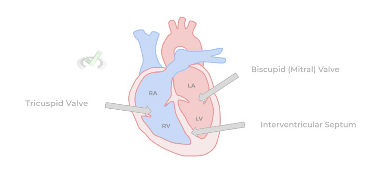
As shown above, the right and left ventricles are separated by the interventricular septum which is basically a continuation of cardiac tissue which extends from the bottom of the heart to separate the ventricular chambers.
The heart also has various valves which help separate the top and bottom chambers — think of valves as kinda like flaps that open and close to allow/prevent the flow of blood from the atria to the ventricles. The right atrium and ventricle are separated by the tricuspid valve while the left atrium and ventricle are separated by the bicuspid (a.k.a. mitral) valve. Both of these valves are called atrioventricular valves because they separate the atria from the ventricles.
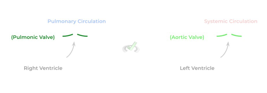
Additionally, the heart also contains valves which separate the ventricles from their respective outflow tracts, termed semilunar valves. Outflow tracts are essentially the tracts which lead into either pulmonary or systemic circulation. Similarly, there are 2 semilunar valves: the pulmonic valve (which separates the right ventricle from pulmonary circulation) and the aortic valve (which separates the left ventricle from systemic circulation).
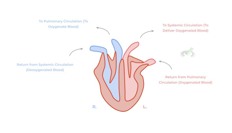
We have a whole other article which goes in much more depth in regards to the flow of blood; however, for know just understand the basic concept that the right side of the heart receives deoxygenated blood which needs to be oxygenated by the lungs in pulmonary circulation.
Conversely, the left side of the heart receives the oxygenated blood from the lungs which is then pumped into systemic circulation to allow for the delivery of blood with all the oxygen and nutrients to systemic tissue.
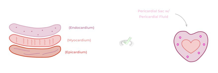
The heart is composed of 3 layers: the epicardium, the myocardium, and the endocardium (from outer to inner most). Additionally, similar to the lung, the heart is surrounded by a fluid filled sac called the pericardial sac which helps to cushion the heart should there be any sudden trauma or movement.
Of great importance are the myocardium and endocardial layers as they play an important part in cardiac contraction. The myocardium provides the actual contractile function of the heart via the presence of cardiac muscle cells (cardiomyocytes).
Both the myocardium and endocardium contain the heart’s electrical conduction system which generates the electrical impulses which triggers muscle contraction. Of special note, these cardiomyocytes are interconnected via gap junctions called intercalated discs to allow for coordinated muscle contraction.
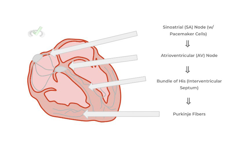
Similar to neurons, cardiomyocytes must have an electrical depolarization which stimulates the cardiac muscle cells to contract. A unique quality about the myocardium is that it’s myogenic, meaning that it can create its own autonomic electrical depolarizations independent of the nervous system.
It accomplishes this via what are called pacemaker cells located in an area of the heart called the sinoatrial (SA) node. These pacemaker cells generate autonomic electrical stimuluses which give the heart its intrinsic rhythm. This electrical stimulus is then propagated throughout the heart to allow for complete cardiac contraction as shown below:
II. Blood Vessels
There are 3 main blood vessels which constitute the cardiovascular system: arteries, veins, and capillaries each of which has a distinct function with regards to the delivery of blood throughout the body.
At its simplest form, arteries carry oxygenated blood AWAY from the heart towards the body’s tissues while veins carry deoxygenated blood TOWARDS the heart in order to be reoxygenated. Capillaries are the smallest divisions of vasculature which are localized within systemic tissue and are the primary site of nutrient and gas exchange.
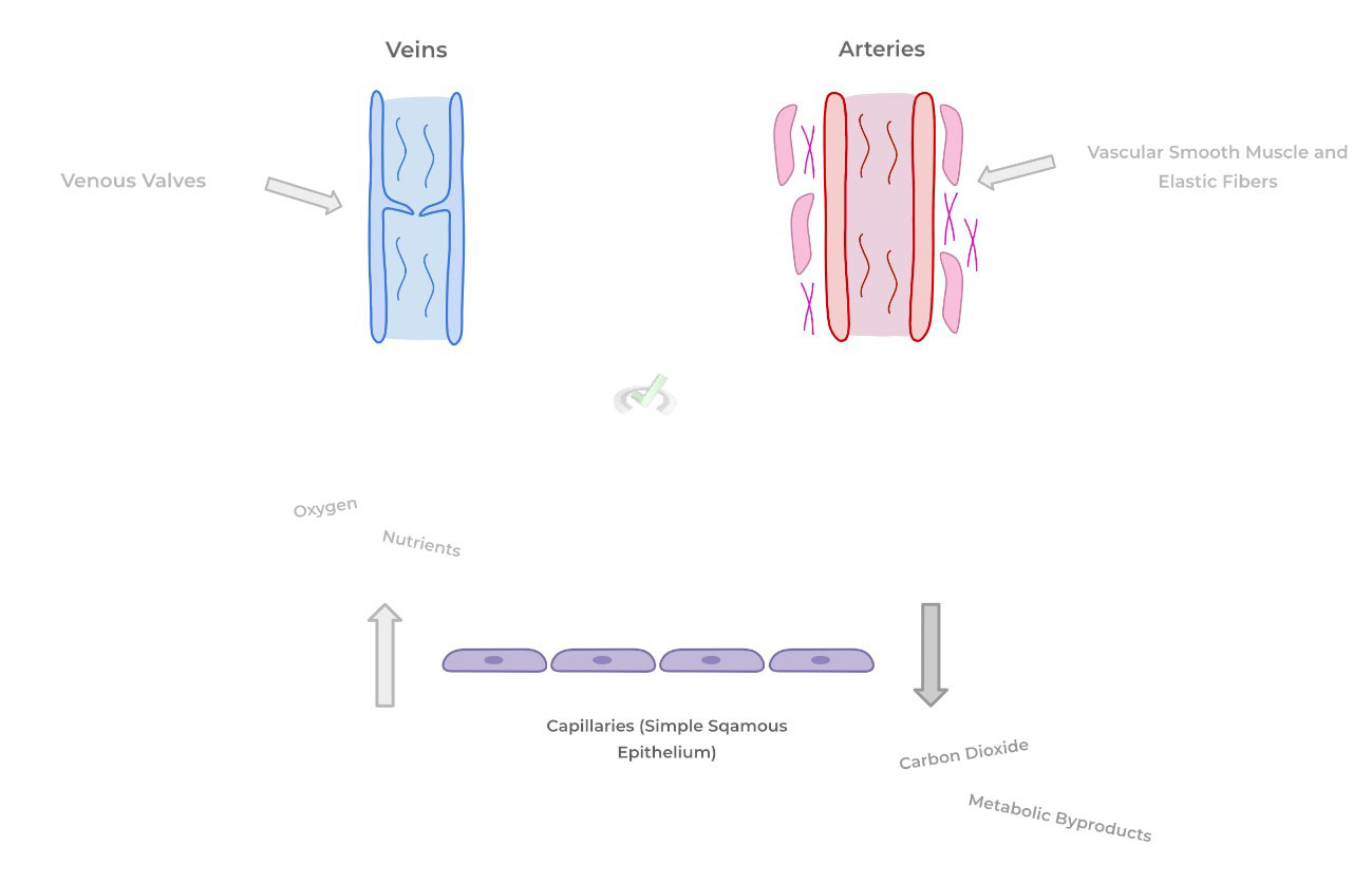
Note the major structural differences between the vasculatures! Arteries generally tend to have abundant smooth muscle and elastic fibers in order to act as one of the main contributors to blood pressure and systemic vascular resistance.
Conversely, while veins aren’t as abundant in smooth muscle and elastic fibers, they contain valves in order to prevent the backflow of blood towards the tissue because blood in the veins must move upwards against gravity to return to the heart!
Finally, recall again that the capillaries are the smallest division of vasculature primarily consisting of a simple, squamous cell lining which facilitates the gas and nutrient exchange at the level of systemic tissue.
III. Blood
Finally, blood is the liquid component of the cardiovascular system that actually travels systematically throughout the body in order to allow for the proper delivery of oxygen and nutrients throughout the body.
Blood can be divided into a cellular component and plasma component. As the name implies, the cellular component contains all the cells within blood that also travel systematically including erythrocytes (red blood cells), immune cells (white blood cells), and platelets (technically, cell fragments).
The plasma component is the actual liquid components and contains other important molecules such as ions, plasma proteins, endocrine hormones, etc.. We have a more in depth covering of the blood components in this article here!
B. Major Structures of the Lymphatic System
Luckily, the structural organization of the lymphatic system is nearly not as complex and primarily consists of the lymphatic vessels and lymph nodes.
Recall that the primary functions of the lymphatic system are to return leaked interstitial fluid back to systemic circulation and to mount an immune response should infection be present!
I. Lymphatic Vessels and Lymph Nodes
An important note about lymphatic vessels is that they participate in ONE WAY circulation as opposed to the two way circulation of the cardiovascular system. The term “one way” comes from the fact that the lymphatic system brings interstitial fluid back into circulation BUT NEVER brings interstitial fluid back towards the tissues.
A great way to think about lymphatic vessels is as a combination of arteries and veins in a sense. Similar to veins, they contain valves to prevent the backflow of lymph and similar to arteries contain vascular smooth muscle which contract to propel the movement of lymph.
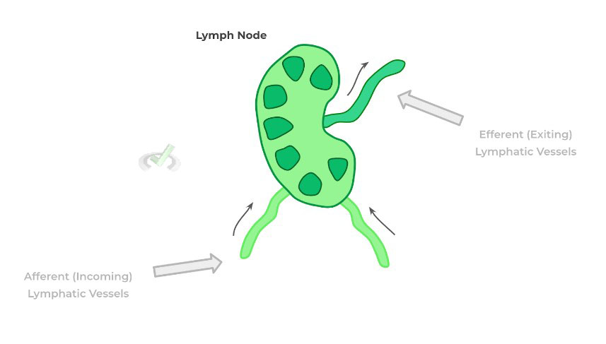
Lymph nodes are bean shaped organs which are present throughout the body and filter incoming lymph entering in from afferent lymphatic vessels for any immunostimulatory antigens. If no antigen is present, the lymph exit via efferent lymphatic vessels and continue their way to return to systemic circulation.
III. Bridge/Overlap
There’s a physiological significance as to why the capillaries are the smallest division of vasculature and is composed of a simple squamous epithelium: this conformation of cells allows for maximum efficiency in regards to maximizing gas and nutrient exchange. To understand this, let’s revisit the concept of diffusion and in particular Fick’s law.
I. Fick’s Law of Diffusion
In its simplest sense, Fick’s law of diffusion states that the flow/flux of a substance across a vessel wall is a product of a permeability coefficient (P), surface area (S), and the concentration gradient (C) divided by the wall thickness (T) as indicated by the equation below:
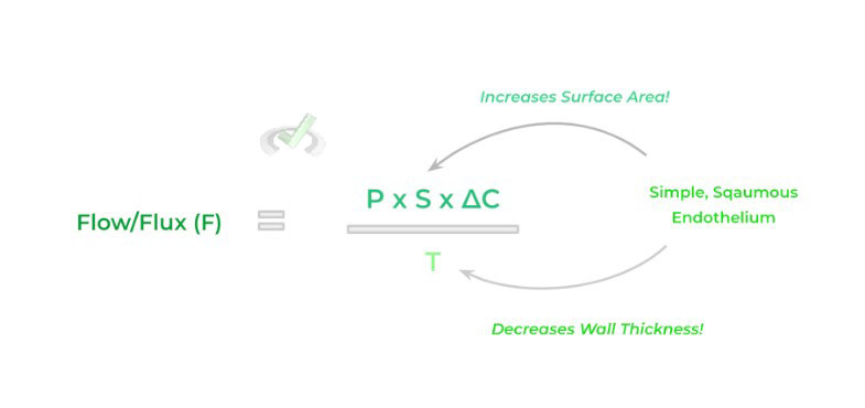
By having a simple squamous epithelium, we can maximize the flow of substances across the capillary wall via increasing the surface area and decreasing the wall thickness! (Be careful! If we decrease the value of the denominator, we’ll increase the flow/flux value).
IV. Wrap Up/Key Terms
Let’s take this time to wrap up & concisely summarize what we covered above in the article!
A. Major Structures of the Cardiovascular System
The 3 main components of the cardiovascular system are the heart, blood vessels, and blood: the heart generates the pumping capacity to allow for the transport of blood throughout the body via blood vessels.
I. Heart
The heart is a four chambered, muscular organ which contains the pumping capacity to drive the flow of blood throughout the body. The heart contains both atrioventricular and semilunar valves which separate the heart chambers and prevent the backflow of blood. Additionally, the heart also contains an interventricular septum, an extension of cardiac tissue from the body which separates the left and right ventricle.
The heart is composed of 3 main layers: the epicardium, myocardium, and the endocardium, all of which is surrounded by the pericardial sac. The heart also contains an autonomic electrical conduction system which provides the internal electrical stimuluses in order to allow for cardiomyocyte depolarizations and subsequent contraction.
II. Blood Vessels
There are generally 3 main types of blood vessels found within the body: arteries, veins, and capillaries. Arteries carry oxygenated blood AWAY from the heart to supply systemic tissue while veins carry deoxygenated blood TOWARDS the heart to be reoxygenated.
Arteries generally have a higher concentration of surrounding vascular smooth muscle and elastic fibers while veins contain valves in order to prevent the backflow of blood into systemic tissue. Finally, capillaries represent the smallest division of blood vasculature as they are composed of simple, squamous epithelium to maximize nutrient and gas exchange.
III. Blood
This constitutes the liquid portion of the cardiovascular system which contains both a cellular component and a plasma component. The cellular components are primarily erythrocytes (red blood cells), immune cells (white blood cells) and platelets (cell fragments for clotting).
The plasma component is the actual liquid component which contains various ions, plasma proteins, and vitamins.
B. Major Structures of the Lymphatic System
Recall that the lymphatic system works synergistically with the cardiovascular and immune system in order to return interstitial fluid to systemic circulation and to mount an immune response should there be an infection present.
I. Lymphatic Vessels and Lymph Nodes
The lymphatic vessels are a one system circulatory system which uptakes leaked interstitial fluid and reabsorbs it back into systemic circulation. Additionally along its pathway, the lymphatic vessels will drain the lymphatic fluid into lymph nodes, bean shaped organs present throughout the lymphatic system which contain abundant immune cells.
Lymphatic fluid enters into the lymph nodes via afferent lymphatic vessels and can present immunostimulatory antigens should an infection be present. Should no infection be present, the lymph exits the lymph node via efferent lymphatic vessels and follow their way back to systemic circulation.
V. Practice
Take a look at these practice questions to see and solidify your understanding!
Sample Practice Question 1
Diastole is the point in the cardiac cycle where there is passive blood flow from the atria to the ventricles to allow for the ventricular filling of blood. Based on this, which combinations of valves should be open during diastole?
A. Tricuspid, Pulmonic
B. Bicuspid, Aortic
C. Tricuspid, Bicuspid
D. Pulmonic, Aortic
Answer: C. Tricuspid, Bicuspid
Sample Practice Question 2
A baby is found to have a congenital cardiac condition called Tetralogy of Fallot. One of the main features of the disease is the presence of a hole within the interventricular septum. What would be the most likely consequence of this malformation?
A. Abnormal mixing of oxygenated and deoxygenated blood
B. Impaired coordinated spread of depolarization
C. Defect in the generation of spontaneous depolarization
D. Reflux of blood from the ventricle into the atrium
Answer: A. Abnormal mixing of oxygenated and deoxygenated blood


 To help you achieve your goal MCAT score, we take turns hosting these