I. What are Eukaryotic Cell TIssues?
A common theme you’ll see in biology is the organization of components into hierarchical structures, where first we start with a big, macroscale view and eventually start getting smaller and more detailed to end up in a microscaled viewpoint.
From complex eukaryotic cells come more complex and high functioning tissues! To make an analogy, if cells are like individual basketball players, then they come together to form the team “tissue”. If the cells had Lebron James, Michael Jordan, Kobe Bryant, Shaq, and Steph Curry, that’d be the GOATED tissue!
Given the analogy above, probably the word “team” is the most important because all the cellular “basketball players” that make up the team tissue must work together in unison to achieve an ultimate goal: in the analogy case: to get the W!!
II. Types of Eukaryotic Cell Tissues
To begin, let’s first define what a tissue is, which is a group of similarly structured cells which come together to perform a function in unison. A step up from this organization are organs!
There are 4 main types of eukaryotic cell tissues: epithelial, connective, muscle, and nervous tissue. We’ll primarily cover the first 2 as the other 2 have their own articles and sections!A. Epithelial Tissue
As implied by the name, these tissues are composed of epithelial cells which line the body’s structure and organs. For example, enterocytes are the intestinal epithelial cells which line the gastrointestinal tract. We’ll come back to this example throughout the article!
I. Structure
The epithelial tissue is composed of 3 main regions: 1) apical, 2) basal, and 3) lateral domain as detailed in the figure below of the gastrointestinal epithelium.
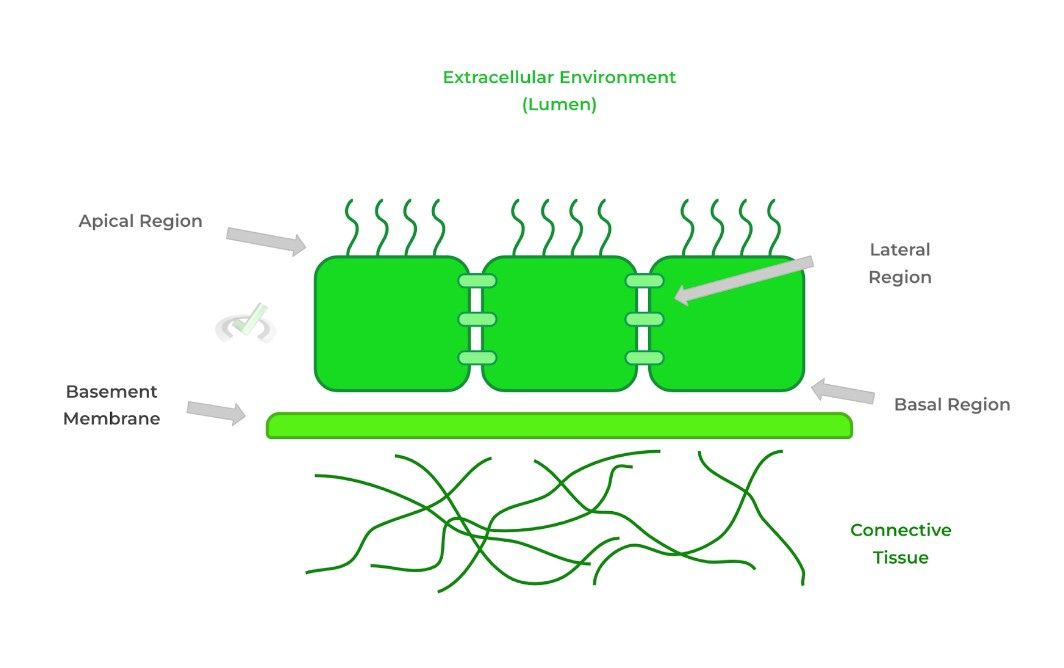
The apical side interacts with the extracellular environment, sometimes called the lumen if the epithelium lines a tube cavity like a blood vessel or the GI tract!
The basal side interacts with the basement membrane, a portion of the extracellular matrix separating the epithelium from underlying connective tissue. The lateral side simply refers to the sides of the cells often connected by cell junctions.II. Function
While its main function is line and serves as a protective barrier, epithelial tissue also has function in the absorption and secretion of substances! Look below for an example!
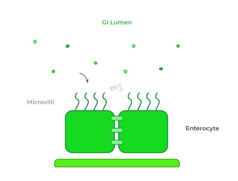
Oftentimes, structures located on the apical side aid in cell function; in this case, the enterocytes’ microvilli aids with the absorption of nutrients.
Most times, the epithelium also constitutes the parenchyma, which is the functional tissue of an organ, even functions aside from the primary ones mentioned above!
For example, hepatocytes are the liver’s epithelial cells, which not only line the liver, but also are responsible for other functions such as detoxification, metabolism, etc.III. Classification by Shape and Layer
Epithelial tissue can be described by the shape of the epithelial cells that compose them which include squamous, columnar, and cuboidal, which are all shaped like they sound like!

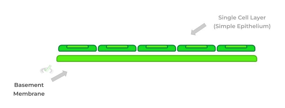
Additionally, epithelial tissue can be described by how their cells are layered. Simple epithelium has only a single layer of cells.
Stratified epithelium is composed of multiple cell layers while pseudostratified epithelial appears to have multiple layers, but are actually composed of one cell layer.
This is due to the single layer having cells of different sizes and types: some small, some tall, some cuboidal, or some columnar!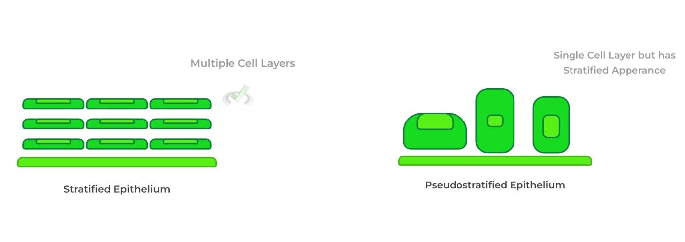
B. Connective Tissue
This type of tissue primarily functions to provide structural support for the body. To again use the previous example, the underlying connective tissue of enterocytes is the lamina propria!
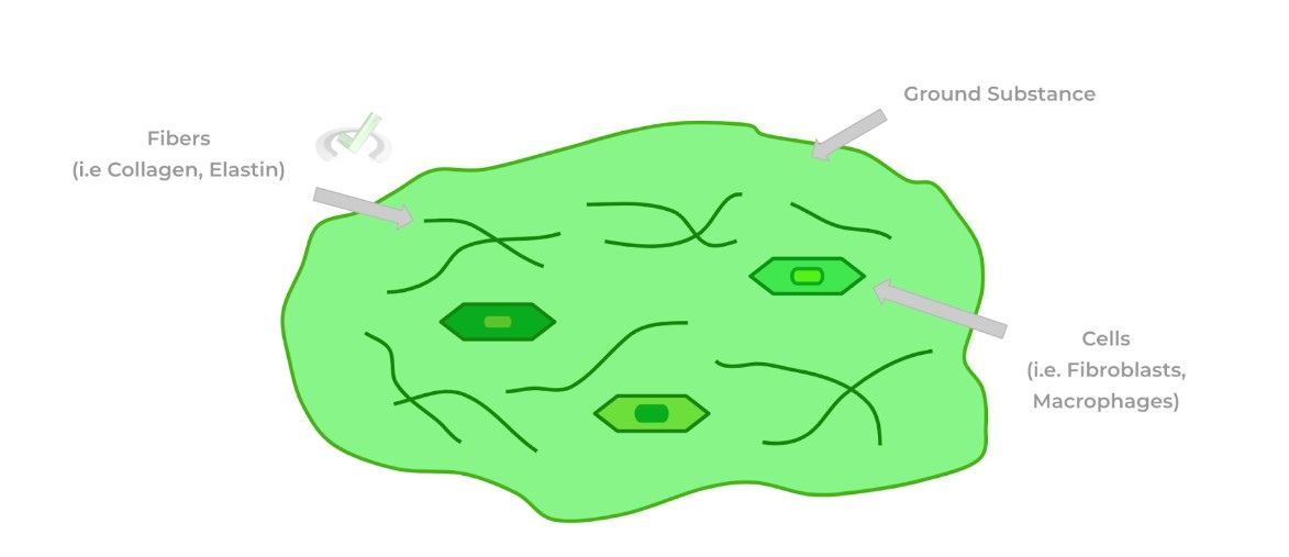
The 3 main components of connective tissue include the ground substance, fibers, and cells. Think of the ground substance as the foundation of the connective tissue described as a gel like substance.
Fibers are strong, flexible proteins embedded within the ground substance which together form the extracellular matrix. Embedded cells serve both structural and immunological roles!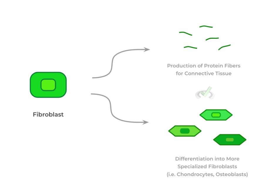
The most prominent type of connective tissue cell is fibroblast, mainly functioning to produce the protein fibers of the connective tissue.
Bones, cartilage, and tendons are all examples of connective tissues! In some of these structures, the fibroblast differentiate into more specialized cells.
For example, fibroblasts in the bone and cartilage differentiate into osteoblasts and chondrocytes, respectively.
Though it may not seem at first, blood is also a connective tissue because it contains a plasma matrix and supports the body through the bodily transport of nutrients and oxygen. It does, however, lack fibers unlike other connective tissue.III. Bridge/Overlap
While the types of cells within a tissue primarily determine its function, the structure and arrangement of cells also play a role in organ function, namely in tissue shape and layer! Let’s take a look at how this plays out!
I. Importance of Tissue Shape and Layer Stratification in Function
It’s best to understand this idea through an example! The function of the alveoli epithelium is to facilitate the transport and exchange of gasses between the alveoli and blood within the capillaries.
It’s crucial that the alveoli epithelium adopts a simple, squamous conformation as this shape and stratification will allow for the most effective transport of gasses.
If the epithelium had a stratified squamous conformation, this would impede gas exchange as the gasses would have a harder time passing through the denser layered epithelium!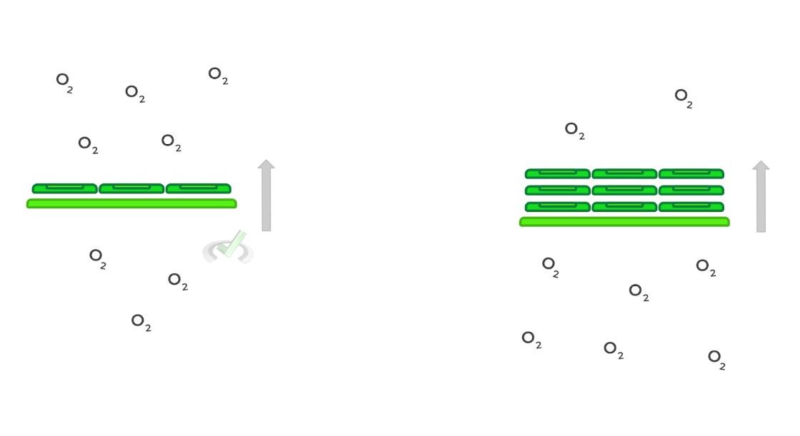
IV. Wrap Up/Key Terms
Let’s take this time to wrap up & concisely summarize what we covered above in the article!
A. Epithelial Tissue
Composed of epithelial cells, the epithelial tissue (epithelium) primarily functions in lining the body’s structure and organs!
I. Structure
The epithelial cells of the epithelium have 3 primary domains: 1) apical, 2) basal, and 3) lateral domain. The apical domain interacts with the extracellular environment, sometimes call the lumen if it lines a tube or vessel.
The basal side interacts with the basement membrane, a portion of the extracellular matrix separating the epithelium from underlying connective tissue.
The lateral side simply refers to the sides of the cells often connected by cell junctions.II. Function
While mainly functioning to line and serve as a protective barrier, epithelial tissue can also function to absorb and secrete substances. Furthermore, structures along the apical side aid in cell function!
The epithelium often also constitutes the parenchyma, which is the functional tissue of an organ, apart from lining, absorption, and secretion.III. Classification by Shape and Layer
Epithelial tissue can be described by the shape of their epithelial cells which include squamous, columnar, and cuboidal.
Additionally, epithelial tissue can be described by how their cells are layered. Simple epithelium has only a single layer of cells.
Stratified epithelium is composed of multiple cell layers while pseudostratified epithelial appears to have multiple layers, but are actually composed of one cell layer.B. Connective Tissue
With its main components being ground substance, fibers, and cells, connective tissue primarily functions to provide structural support for the body.
The ground substance acts as the main gel-like foundation embedded with strong, flexible protein fibers and cells which have structural and immunological function.
Fibroblasts are the most abundant connective tissue cells which synthesize and release the protein fibers and can differentiate into more specialized cells in different connective tissues such as bones, cartilage, and tendons.V. Practice
Take a look at these practice questions to see and solidify your understanding!
Sample Practice Question 1:
The epithelial enterocytes within the small intestine also contain some of the main disaccharidases like lactase and sucrase. Which domain would these enzymes be located?
A. Apical
B. Basal
C. Lateral
D. Basal and Lateral
Ans. A
Recall the apical side of the epithelium interacts with the extracellular environment or lumen. Thus, it’d be logical for these enzymes to also be located within the apical domain in order to digest the incoming disaccharides.
Specifically, these disaccharidases lie within the microvilli of the enterocytes, which increases the surface area for digestion!
Sample Practice Question 2:
GIven the following epithelial tissue characteristics, which of the following should describe the characterization of the endothelial cells which line the capillaries?
I. Stratified
II. Simple
III. Pseudostratified
IV. Squamous
V. Cuboidal
VII. Columna
A. I and IV
B. II and VI
C. III and V
D. II and IV
Ans. D
Think back to the function of capillaries! These are the thinnest divisions of blood vessels because this is where the exchange of gasses, nutrients, and other substances occurs between the blood and tissue.
As such, a simple, squamous epithelial configuration will be the most effective in their transportation as this is the least dense cellular conformation.


 To help you achieve your goal MCAT score, we take turns hosting these

