I. What are the Structural and Physiological Characteristics of Prokaryotic Cells?
Having appeared much earlier along evolutionary lineage, it comes to no surprise that when compared to their more complex eukaryotic counterparts that prokaryotes are much similar in both structure and their cellular processes!
The study of prokaryotic cell structure has been revolutionary in biology and medicine not just in terms of developing our scientific curiosity, but also in utilizing these understandings to develop medicinal therapeutics against their infections.
When reviewing this section, a good way to internalize and really understand the information is to make a side by side comparison and contrast to the structural characteristics of eukaryotes! One great way to do this is through generating a table!
II. Features Common in Prokaryotic Cells
Though they all share common components, we’ll approach discussing this article through focusing on bacteria. Note that archaea also shares these common features with very few and minor differences!
A. Lack of Nucleus and Mitotic Features
One of the most defining features of prokaryotes that differentiates them from eukaryotes is their lack of a true nucleus.
As opposed to a true nucleus enclosed by a nuclear membrane, the bacteria’s circular DNA chromosome is located in a region called the nucleoid, which is basically the free space the bacterial DNA occupies.

Additionally, due to their lack of true nucleus, prokaryotes also lack several of the mitotic features found in eukaryotic mitosis such as the spindle apparatus and centrioles.
Rather, prokaryotes participate in a much simpler form of reproduction in the form of binary fission, as covered in another article!B. Lack of Membrane Bound Organelles
Another distinct feature of prokaryotes differentiating them from eukaryotes is their lack of membrane bound organelles, including mitochondria, endoplasmic reticulum, etc.
However, it must be remembered that prokaryotes still have ribosomes, as without them, prokaryotes would be able to synthesize the necessary proteins and enzymes!
Note that the prokaryotic ribosomes are a little smaller than those found in eukaryotes, as listed by their subunit components below.
Another good point to make is that because they lack mitochondria, the electron transport chain in prokaryotes is found in their cell membrane!

You’ll notice that a lot of metabolic processes remain the same between the prokaryotes and eukaryotes, but just take place in different places!
To give another example, the Krebs Cycle (this again!?) takes place in the cytoplasm in prokaryotes due to the lack again of mitochondria!C. Presence of Bacterial Cell Wall and Composition
Another important bacterial structure is the cell wall. While there are eukaryotes that do have cell walls, this is not the case for our human animal cells.
While there are many components to the cell wall, the most important is peptidoglycan, which is a type of polysaccharide.
It’s when the multiple polysaccharides are cross-linked or bonded together by the enzyme transpeptidase that structural rigidity and strength is given to the cell!
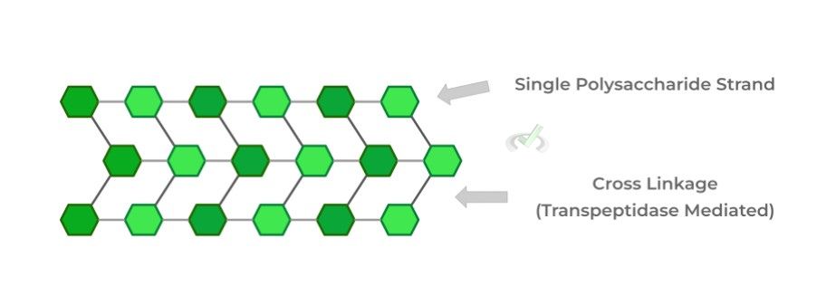
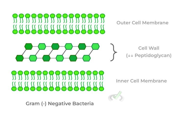
Bacterial cells can also be categorized into Gram positive (+) or negative (-) bacteria which differ in the amount of cell wall peptidoglycan and the arrangement of the cell wall and membrane.
Gram (+) will have a high amount of peptidoglycan in the cell wall which surrounds the cell membrane and will appear color purple in a gram stain.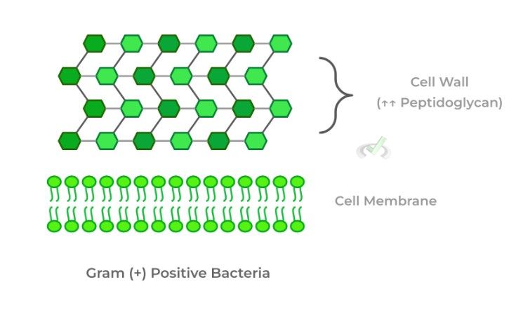
Gram (-) will have a low amount of peptidoglycan but is actually “sandwiched” between an outer and inner cell membrane. When stained, the bacteria will appear red/pink!
D. Flagella Structure and Propulsion in Chemotaxis
Additionally, prokaryotes can also produce locomotion utilizing their flagella which is a tail-like projection made from flagellin protein and causes movement of the cell through its rotation!
Its 3 main components are the filament, hook, and basal body. The basal body is responsible for generating the rotation needed for movement.
The hook acts as a bridge between the filament and basal body so that when the basal body rotates, the filament rotates as well, generating movement.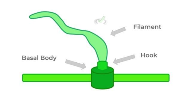
A key use of the flagella is when the bacterial cell needs to move in chemotaxis, which is the movement of a cell in response to a chemical stimulus.
This is important for prokaryotes, especially being unicellular, as these chemical stimuli often indicate a source of energy the cell can use while also moving away from a toxic one.
E. Symbiotic Relationships with Other Species
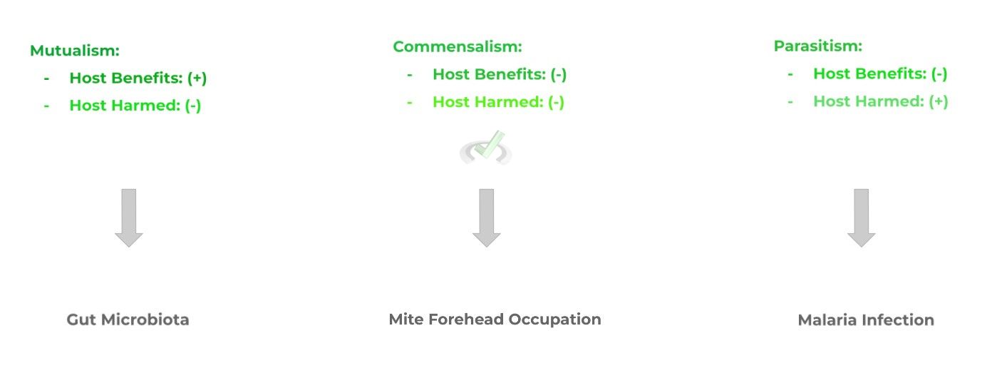
Oftentimes, bacterial cells live with another species, usually a larger host species. This shared living arrangement interaction between the 2 is termed symbiosis.
Certain relationships can form between the species and differ in the benefits and harm (or lack thereof) that the host species experience. In all the scenarios, however, the bacteria always benefits. Look at the ones listed below!
III. Bridge/Overlap
Let’s talk and explain a little more about the example given above for a mutualistic relationship as it’s a really interesting one that’s evolved between bacteria and humans!
I. Mutualism between Bacteria and the Human GI Tract
An interesting topic of discussion in past years has been the effects of the human microbiota and its impact on human health!
Bacterial cells obviously benefit by having an area to live in the intestines while also being supplied with nutrients from human food consumption.
Humans too benefit as the bacterial cells not only sometimes aid in digestion but also have an immunological role by preventing the colonization of harmful pathogens!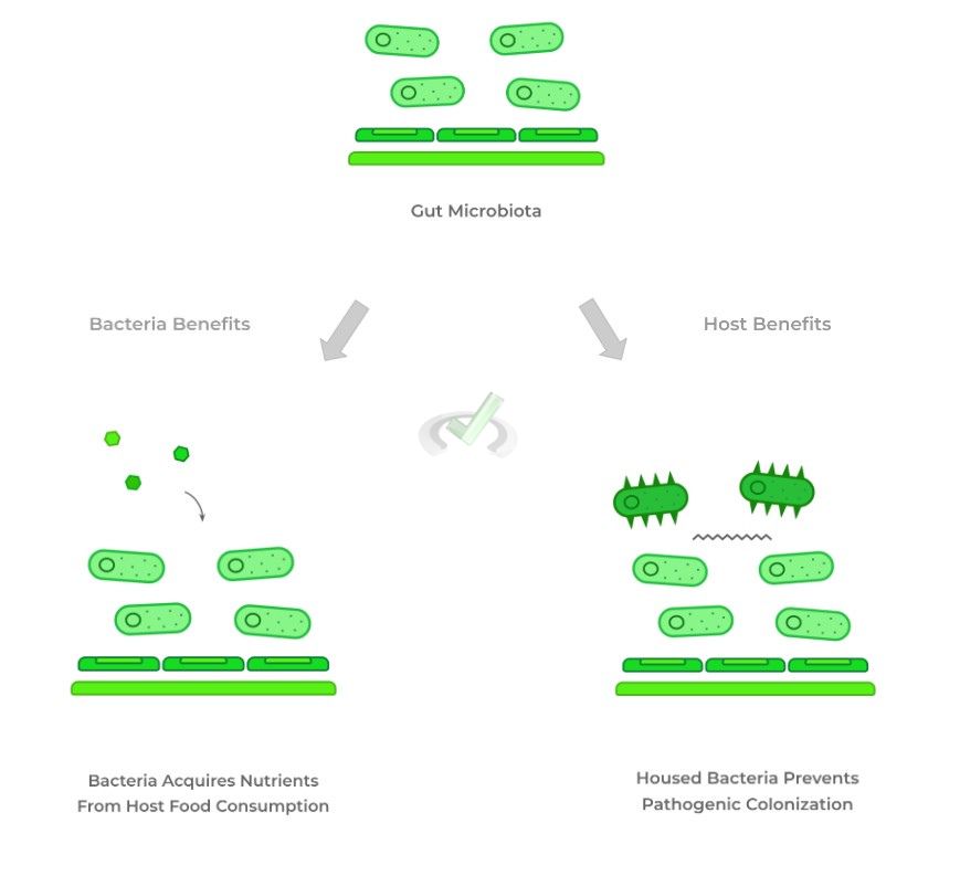
IV. Wrap Up/Key Terms
Let’s take this time to wrap up & concisely summarize what we covered above in the article!
A. Lack of Nucleus and Mitotic Features
The lack of a true nucleus is one of the defining features of prokaryotes as their circular DNA chromosome is housed in a region called the nucleoid.
Additionally, prokaryotes lack the mitotic features such as the spindle apparatus and centrioles as seen in eukaryotes and rather participate in binary fission.B. Lack of Membrane Bound Organelles
Similarly, the absence of membrane bound organelles such as the mitochondria and endoplasmic reticulum is another defining feature of prokaryotes. However, they still contain ribosomes!
Prokaryotes have a slightly smaller ribosome, consisting of 30s/50s subunits for a 70s ribosome. Eukaryotic ribosomes have 40s/60s subunits for a 80s ribosome!
Another important note to make is that because they lack mitochondria, the electron transport chain is found within the prokaryotic cell membrane!C. Presence of Bacterial Cell Wall and Composition
Unlike human eukaryotic cells, bacterial cells contain a cell wall composed of multiple peptidoglycan cross-linkages, catalyzed by transpeptidase!
Gram (+) positive bacteria have a high amount of peptidoglycan in their cell wall surrounding the cell membrane. When gram stained, the bacteria will appear purple.
Gram (-) negative bacteria have a low amount of peptidoglycan in their cell wall but are “sandwiched” between an outer and inner cell membrane. When gram stained, the bacteria will appear red/pink.D. Flagella Structure and Propulsion in Chemotaxis
Composed of flagellin protein, the flagella is a tail-like projection that’s involved in bacterial locomotion, composed of 3 main components: filament, hook, and basal body.
The basal body is responsible for generating the rotation needed for movement, while the hook acts as a bridge between the filament and basal body which allows for rotation of the filament when the basal body rotates.
Bacterial locomotion is particularly important in chemotaxis, which is the movement of a cell in response to a chemical stimulus.E. Symbiotic Relationships with Other Species
Different symbiotic relationships can emerge between bacteria and the host species they’re inhabiting and differ on whether they cause benefit or harm to the host.
Mutualism occurs when both the bacteria and host benefits from one another.Commensalism occurs when only the bacteria benefits, but the host is not harmed.
Parasitism is where the bacteria benefits but causes harm to the host species.
V. Practice
Take a look at these practice questions to see and solidify your understanding!
Sample Practice Question 1:
Oftentime, medicinal therapeutics have to target differences between bacteria and human cells so that only the bacterial cells will be affected. Which of the following could be utilized as targets?
I. Ribosome Structure
II. Transpeptidase
III. Cell Membrane Disruption
A. I only
B. III only
C. I and II
D. I and III
Ans. C
Recall that prokaryotes have a slightly smaller 70s ribosome structure composed of 30s/50s subunits when compared to eukaryotes. Because of this difference antibiotics can actually specifically target and inhibit the 70s ribosomes of prokaryotes.
Additionally, only bacteria will have a cell wall compared to human cells! As such, targeting transpeptidase, the enzyme which catalyzes peptidoglycan crosslinking, will specifically disrupt the formation of the cell wall in the bacteria.
Sample Practice Question 2:
Given the gram stain shown below, what can be said about the bacteria that appears on the gram stain?

A. The bacteria has a high peptidoglycan concentration in its cell membrane
B. The bacteria has a low peptidoglycan concentration in its cell wall
C. The bacteria has its cell wall sandwiched between an outer and inner cell membrane
D. The bacteria has a high peptidoglycan concentration in its cell wall
Ans. A
Watch the wording! A purple gram staining indicates a Gram (+) positive bacteria, characterized by a high peptidoglycan concentration in its cell wall which surrounds the cell membrane.



 To help you achieve your goal MCAT score, we take turns hosting these