I. What are the Structures and Processes of Mitosis?
What did one cell tell to her sister when she stepped on her toe? “Mi-to-sis”. Alright, now that we got that classic, albeit corny, joke out of the way, it’s no surprise how important this topic is not just to the MCAT but in basic cellular biology.
Essentially the eukaryotic equivalent of binary fission, mitosis too allows for the generation of 2 daughter cells. But being a eukaryotic cell feature, it’s definitely a bit more complex than its prokaryotic counterpart as it comes with its own intricacies and complexities.
As with many topics on the MCAT, we feel the best way to approach studying mitosis is through using visuals to detail each step along the pathway. We’ll for sure include these visuals along the article but also drawing them for yourself might be a useful study tool!
II. Stages of Mitosis
Similar to the cell cycle, mitosis is made up of 4 main phases: prophase, metaphase, anaphase, and telophase. Sometimes, you might see cytokinesis squeezed in with telophase and other times they might be separate.
A. Prophase
After passage from the G2/M checkpoint, prophase occurs and is characterized by nuclear envelope breakdown as well as chromatin condensation into chromosomes each with 2 sister chromatids.
The condensation is important as it facilitates easier separation of the duplicated DNA during anaphase as we’ll cover soon!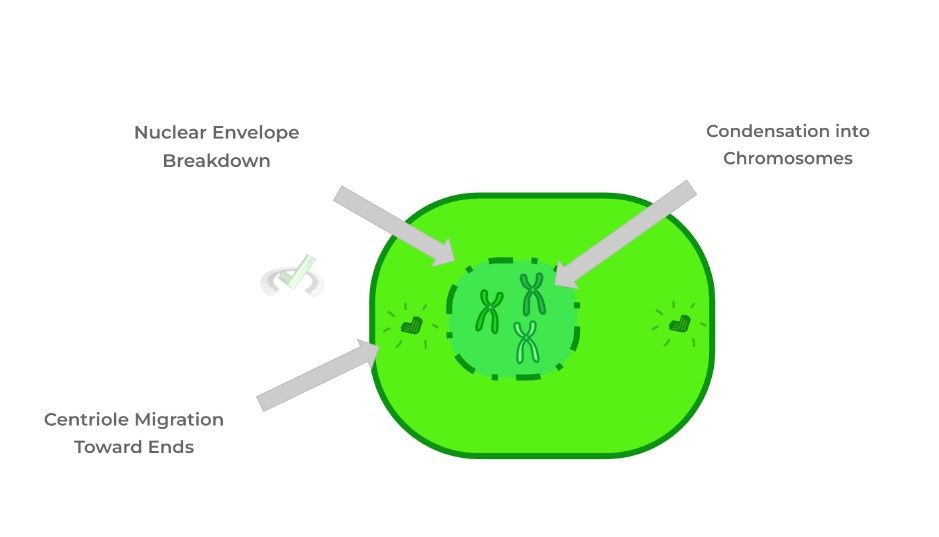
I. Mitotic Structure: Centrioles and Centrosome
Another staple of prophase is the migration of centrioles towards the opposite ends of the cells to form what’s called the centrosome. Centrioles are cylindrical shaped organelles composed of microtubules, specifically tubulin.
The centrosome is the main microtubule organizing center and can be thought of as the region where 2 centrioles at right angles reside while organizing the spindle fibers as we’ll discuss.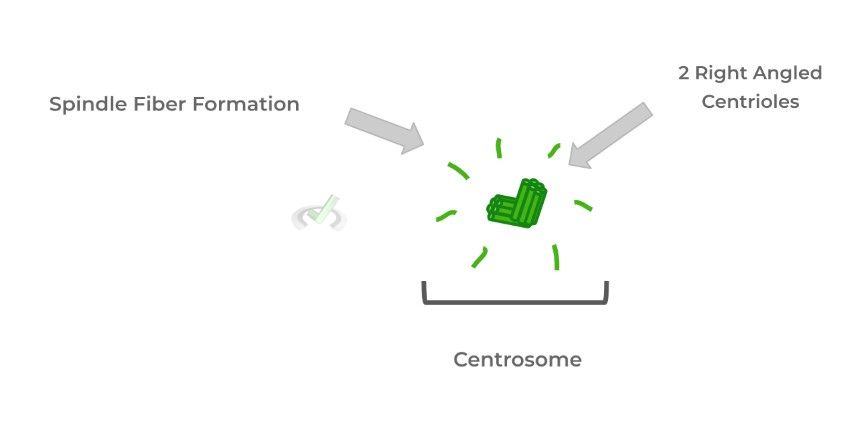
B. Metaphase
With the DNA now condensed into chromosomes with the 2 sister chromatids, the chromosomes then aligned along the equator/midline of the dividing cell forming what’s called the metaphase plate.
As shown in the figure below, the sister chromatids are arranged where each one faces a different end which is important for separation during anaphase.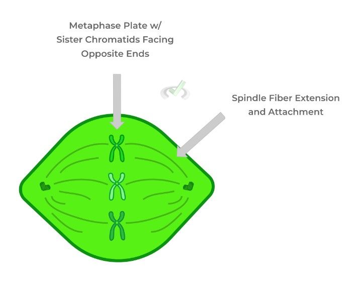
I. Mitotic Structure: Spindle Fibers
As mentioned before, the spindle fibers are generated by the centrioles of the centrosome and begin to “extend” and grow out from the centrosome eventually attaching to the kinetochore of the centromere, as we’ll discuss in a few.
Just like the centrioles, the spindle fibers are composed of the microtubule protein, tubulin and just like other microtubules, they lengthen or shorten via the addition or removal of tubulin subunits from the microtubule ends, respectively.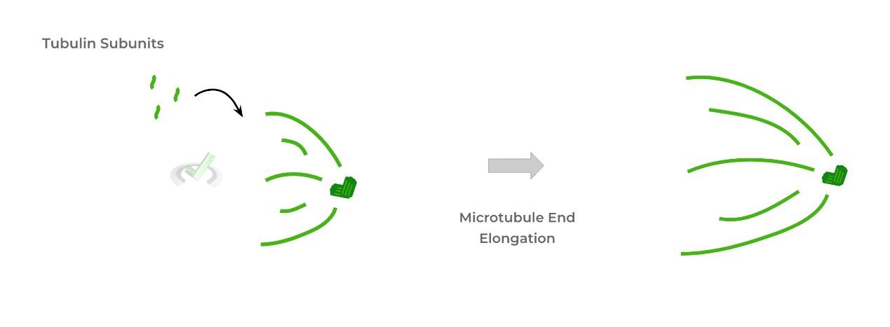
C. Anaphase
After attachment of the spindle fibers to the centromere’s kinetochore, the spindle fibers begin to retract which separates the sister chromatids as each one moves toward opposite ends of the cell.
Now when the cell divides down the midline to make 2 daughter cells, each daughter cell contains the same amount of DNA as its parent cell.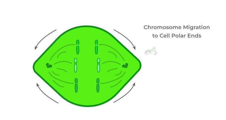
I. Mitotic Structure: Kinetochore
Recall that the centromere is the region of the chromosome which holds the 2 sister chromatids together. This region is actually composed of many proteins which function in mitotic division, one of which is kinetochore.
Think of the kinetochore proteins as like docking points for the spindle fibers to attach to so that when they retract, the chromatids are “pulled” apart.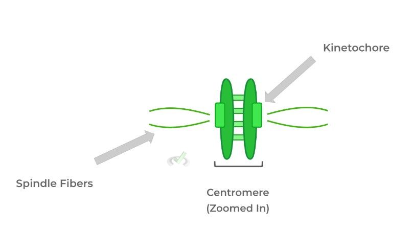
D. Telophase (and Cytokinesis)
In this final phase, we essentially see a reversal of the events that occur in prophase. These include the disappearance of the spindle fibers, the reforming of the nuclear envelope, and the decondensation of the chromosome to the more “free-flowing” chromatin structure.
In cytokinesis, contractile actin rings organize in the midline which contract and “pinch” the cytoplasm of the cell until 2 daughter cells are separated and formed. The indentation created as shown below is called the cleavage furrow.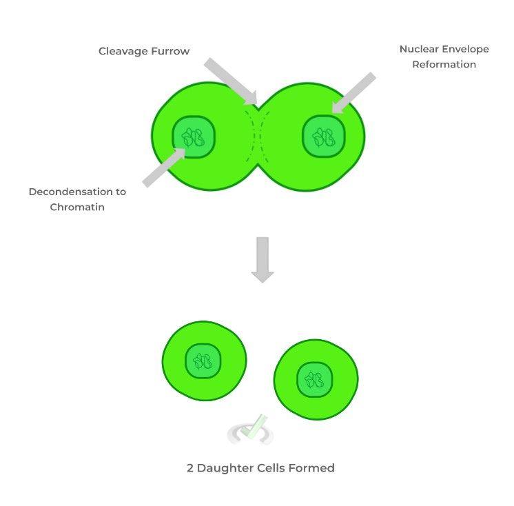
III. Bridge/Overlap
It can be a little confusing understanding which is a chromatid and which is a chromosome. That being said, let's try to get a better understanding of how and when to use the terms!
I. Terminology Clear Up: Chromosome v.s. Chromatid
When the term chromosome is used, it generally refers to DNA in its most condensed form most prominent during mitosis. Chromatids basically refer to the 2 individual, duplicated strands of DNA generated during the S phase of the cell cycle.
Sometimes, however, the term chromosome can also refer to the 2 sister chromatids held together by the centromere.
This is especially the case in metaphase when the chromosomes (with both the sister chromatids) align in the metaphase plate.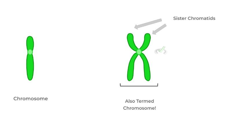
IV. Wrap Up/Key Terms
Let’s take this time to wrap up & concisely summarize what we covered above in the article!
A. Prophase
After passage through the G2/M checkpoint, the nuclear envelope begins to degrade in order to allow for the condensed chromosomes to begin to migrate toward the midline of the cell.
The condensed form of the chromosomes allows for easier separation of the sister chromatids during anaphase.I. Mitotic Structure: Centrioles and Centrosome
Additionally, prophase marks the migration of centrioles to the polar ends of the cell where they organize into regions called centromeres which function as the main microtubule organizing center.
Centrosomes are composed of 2, right-angled centriole organelles which are composed of tubulin and will work to generate the spindle fibers needed in metaphase and anaphase.B. Metaphase
The condensed chromosomes, now released from the nuclear envelope, migrate and align at the midline/equator of the cell, with this alignment being called the metaphase plate.
In addition, the chromosomes are oriented in a way that each chromatid faces opposite sides of the cell so that when they separate, each cell receives an equal amount of DNA.I. Mitotic Structure: Spindle Fibers
The centrioles within the centrosome begin making spindle fibers, which are also microtubules made of tubulin which extend out and attach to the kinetochores of centromeres.
C. Anaphase
The spindle fibers attached to the kinetochore begin to “retract” and decrease in length which causes the separation of the 2 sister chromatids.
From this separation, each daughter cell will have the same amount of DNA as its parent cell.I. Mitotic Structure: Kinetochore
Within the centromere, the region holding the 2 chromatids together, is the kinetochore which is a protein which serves as a docking point for spindle fibers to attach to.
D. Telophase (and Cytokinesis)
In telophase, we basically see a reversal of the events in prophase: the nuclear envelope reforms, the chromosomes decondense back to chromatin, and the spindle fibers disappear.
In addition, contractile actin rings form around the cytoplasm and begin “pinching” them and creating an indentation called the cleavage furrow. Eventually, this “pinching” results in the cell splitting, generating 2 daughter cells.
V. Practice
Take a look at these practice questions to see and solidify your understanding!
Sample Practice Question 1:
Which of the following lists the mitotic events below in order?
I. Nuclear Envelope Reforms
II. Metaphase Plate Forms
III. Spindle Fibers Begin to Retract
IV. Condensation of Chromatin to Chromosomes
A. I, II, III, IV
B. II, III, I, IV
C. IV, II, III, I
D. II, IV, I, III
Ans. C
This answer choice follows the process of mitosis, which involves prophase (condensation to chromosomes), metaphase (metaphase plate formation), anaphase, (spindle retraction), and telophase (nuclear reforms).
Sample Practice Question 2:
Suppose a parent cell has 3 chromosomes. Upon one round of mitotic division, however, it’s found that one daughter cell has 4 chromosomes while the other daughter cell has 2. Which phase of mitosis most likely could have been defective which resulted in this abnormality?
A. Prophase
B. Metaphase
C. Anaphase
D. Telophase
Ans. C
Recall that anaphase is the phase of mitosis where the sister chromatids separate in order to evenly distribute the chromatids and create 2 genetically identical daughter cells.
However, if separation does not occur, one cell could end up with both sister chromatids instead of one resulting in an extra acquired chromosome. This process if actually called disjunction and is present in many diseases.







 To help you achieve your goal MCAT score, we take turns hosting these




 reviews on TrustPilot