I. What is the Hierarchical Structure of Muscle Tissue?
It’s a popular saying among military generals that battles and wars are one by structure and hierarchy — though it may not be as serious as when applied to the physiological systems like the muscular system, the theme of structure and hierarchy are important in regards to understanding the muscle function.
As we’ll get into, it’s amazing to see the various breakdowns of the muscle from the largest macroscale towards the miniscule microscale. With that be said, let’s go ahead and see how the muscle tissue is broken down into its various components!
II. Muscle Tissue From the Macro to the Microscale
When breaking down the hierarchical structure of the muscle, we’ll primarily break it down into 2 parts: 1) from the muscle to the cellular component and then the 2) muscle cell component to the organelle component. These will all make a little more sense when we cover them in a stepwise fashion.
A. Muscle to Muscle Cell
The summative collection of all the underlying hierarchical structures (as we’ll touch on soon) yields a single muscle tissue: think of the biceps, triceps, quadriceps, etc. From here, we have the first subdivision from the muscle which are fascicles.
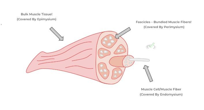
Fascicles are essentially bundles of muscle cells which are clumped together. When multiple fascicles come together, they then form the bulk muscle. Finally, the muscle cells (also called muscle fibers) are the individual cells which contain the contractile organelles to initiate contraction.
Additionally, there are important surrounding connective tissue which surrounds each of these 3 different hierarchical muscle structures which add an extra component of support and separation of each hierarchical component while also serving as the nerve and blood supply to the muscle as shown below!

‘As shown above, the epimysium is the connective tissue sheath covering the bulk muscle tissue, the perimysium covers the muscle fascicles, and the endomysium encloses individual muscle cells (also called muscle fibers).
B. Muscle Cell to Contractile Components
As we mentioned above, muscle fibers are the individual cell components which comprise muscle and contain the individual contractile organelles. Within each muscle cell are specialized contractile organelles called myofibrils!
To break it down even further, the myofibrils contain numerous amounts of sarcomeres which are the actual contractile elements that initiate muscle contraction. This is due to the actin and myosin proteins present within sarcomeres which engage in a process called cross bridge cycling to allow for muscle contraction as we’ll describe in further detail in another article!

As shown above, each smaller and microscale component all come together collectively to form the bulk muscle tissue!
While again we don’t like to play favorites in regards to what we want to focus on, the MCAT tends to focus on content and questions at the level of the sarcomere because of their subcellular processes which eventually leads to muscle contractions!
III. Bridge/Overlap
While in this article we’ve covered the hierarchical structure of the muscle tissue, it’s amazing to see that this hierarchical pattern is also seen in other organ systems throughout the body! Let’s go ahead and give you all an example while relating it to its muscular counterparts!
I. Analogous Hierarchical Organization of Peripheral Nerve Tissue
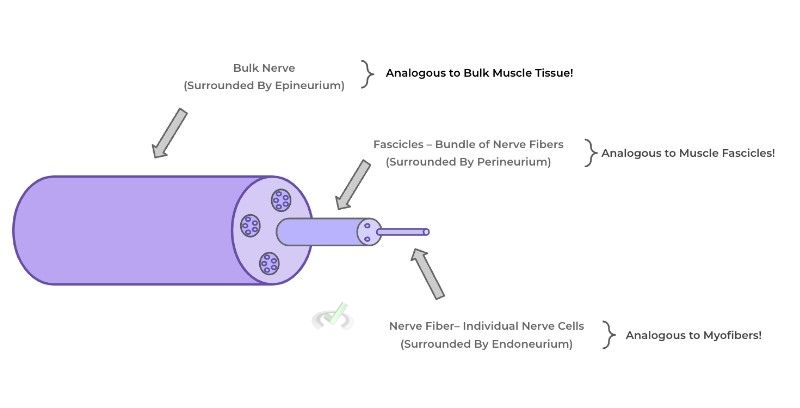
It’s really cool to note that the peripheral nerves also have a similar hierarchical organization structure with that of muscles! The bulk nerves are essentially analogous to the bulk muscle — composing these nerves are bundles for nerve fibers (which are the individual nerve cells/neurons) also arranged in fascicles!
Additionally, each of the nerve components is also wrapped by a sheath of connective tissue similar to its muscular counterparts: the bulk nerve is covered by the epineurium, the fascicles are covered by the perineurium, and the individual nerve fibers are covered by the endoneurium! (Notice the similarities in the prefixes!).

IV. Wrap Up/Key Terms
Let’s take this time to wrap up & concisely summarize what we covered above in the article!
A. Muscle to Muscle Cell
The bulk muscle tissue is composed of a collection of fascicles which are essentially bundles of muscle fibers. Muscle fibers are the actual individual muscle cells which contain the organelles which will allow for contraction!
These muscular components are also wrapped in their respective connective tissue sheaths which add an extra component of support while also being a supply of blood vessels and nerves. These include the epimysium (which covers the bulk muscle), the perimysium (which covers the fascicles), and the endomysium (which covers the individual muscle cells).
B. Muscle Cell to Contractile Component
As mentioned above, the individual muscle cells contain the actual contractile rod-shaped organelles called myofibrils! Likewise, these myofibrils contain the actual contractile elements, called sarcomeres, which allow for muscle contraction due to a process called cross bridge cycling of actin and myosin proteins!
V. Practice
Take a look at these practice questions to see and solidify your understanding!
Sample Practice Question 1
Complete the statement.
______________ are bundles of _____________. These bundles are enclosed by a sheath of connective tissue called ____________.
I. Myofibrils
II. Fascicles
III. Myofibers
IV. Epimysium
V. Perimysium
VI. Endomysium
A. II, III, V
B. III, II, VI
C. I, II, IV
D. II, I, V
Ans. A
Recall that fascicles are essentially bundles of myofibers, which are the individual muscle cells — the connective tissue which envelopes and covers the fascicles is called perimysium. The epimysium and the endomysium encapsulate the bulk muscle tissue and individual muscle cell/myofibers, respectively.
Sample Practice Question 2
Polymyositis is a chronic inflammatory disease which is characterized by inflammation of the muscle. Recent research has shown that it primarily involves inflammation of the endomysial layer of the skeletal muscle. Which hierarchical muscular component would be most directly affected as a result?
A. Bulk Muscle Tissue
B. Fascicle
C. Sarcomeres
D. Myofibers
Ans. D
Recall that myofibers refer to the individual muscle cell and are wrapped by a sheath of connective tissue called endomysium. As such the hierarchical muscle structure which most likely would be affected is the myofibril/muscle cell.


 To help you achieve your goal MCAT score, we take turns hosting these


 reviews on TrustPilot