I. What is an Action Potential and How Is It Propagated in the Nervous System?
While we primarily look at the functioning body from a biological standpoint, it’s pretty amazing to realize that our physiology of our body relies just as much on electrochemistry to properly function — look no further than how neurons communicate with one another.
While you probably won’t have to calculate membrane voltages and use all these fancy equations (unfortunately, these tasks are more required for the Chem/Phys section of the MCAT), we’ll definitely need to apply some basic electrochemistry to see how it results in neurons communicating with one another!
II. The Action Potential
We’ll divide the discussion of the action potential into 2 main parts: 1) a larger scale overview of action potential propagation down the neuron and 2) the voltage changes that occur which allow for the propagation.
A. Macroscale Propagation of the Action Potential
An action potential is essentially a signal that can be generated in a neuron due to changes in the membrane voltage and subsequently propagated down the neuron to allow for the release of neurotransmitters.
Let’s first give a little spatial context: recall that the synapse is a communicating junction between 2 neurons. Composing a synapse are a presynaptic neuron (the neuron which releases the neurotransmitters from the axon terminal) and a postsynaptic neuron which binds to the released neurotransmitters via receptors on dendrites.
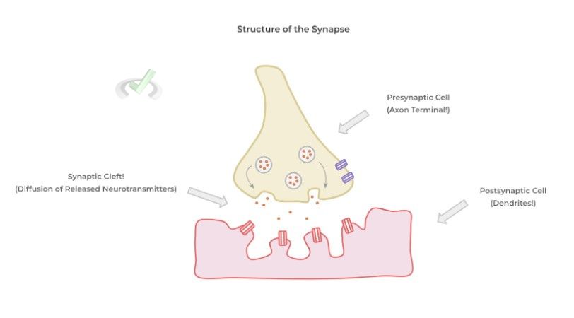
After receptor binding, the effects of the excitatory and inhibitory neurotransmitters are summated – if the excitatory stimuli outweighs the inhibitory, an action potential is generated!
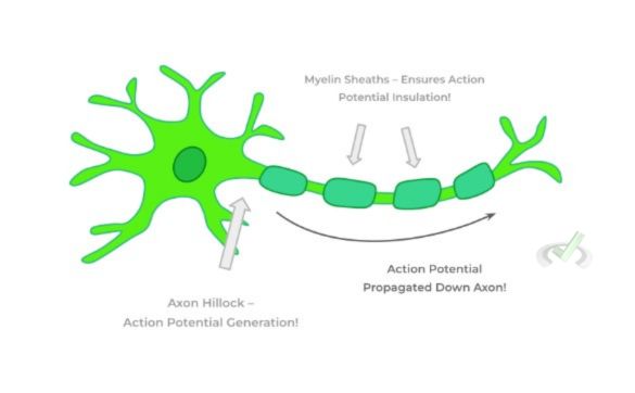
The action potential is generated in a region called the axon hillock (where the cell body meets the axon) and is propagated down the axon.
Note also the presence of myelin sheaths covering the axon which aids in insulating the action potential to ensure that the electrical signal is propagated at a sufficient speed and by the time it arrives at the axon terminal to complete the process. strength
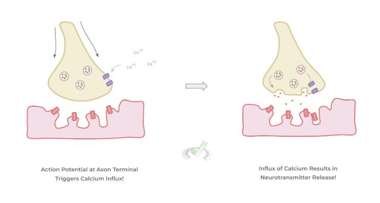
When the action potential reaches the axon terminal, this triggers the influx of extracellular calcium into the cell. This is an important event because the influx of calcium triggers the fusion of the vesicles containing neurotransmitters with the membrane
This results in the release of the neurotransmitters into the synaptic cleft, which can then bind to postsynaptic receptors and eventually repeat the cycle.
B. Action Potential Generation
As mentioned before, the generation of an action potential results from changes in membrane potential due to changes in the influx/efflux of various ions. Let’s go ahead and breakdown the various stages of action potential.
I. Neurotransmitter Summation and Stimulus
In this stage, we’re simply seeing if the summated neurotransmitters can depolarize (i.e. make the membrane potential more positive) the membrane enough in order to trigger the action potential.
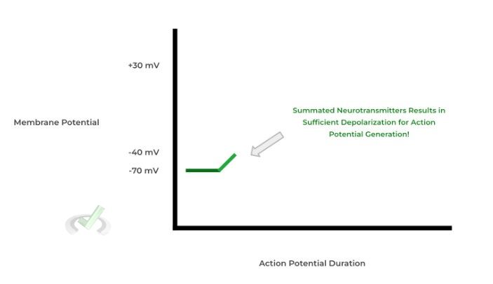
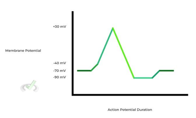
With the resting membrane potential of the neuron being about -70 mV, neurotransmitter receptor binding results in the activation of ligand gated ion channels which allows for influx of ions.
If the influx ions result in enough initial depolarization – usually, increase the membrane potential from -70 mV to -40 mV — an action potential is triggered!
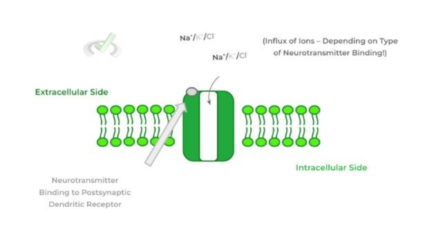
As mentioned, when neurotransmitters pass to the dendritic receptors, they activate specific ligand gated ion channels which allow for the influx of certain ions.
These ions can either further depolarize the membrane to reach the action potential threshold or further hyperpolarize (i.e. make more negative) which makes them less likely to generate an action potential.
Excitatory neurotransmitters generally result in the influx of positive ions (cations) which further depolarize the membrane while inhibitory neurotransmitters result in the influx of negative ions!
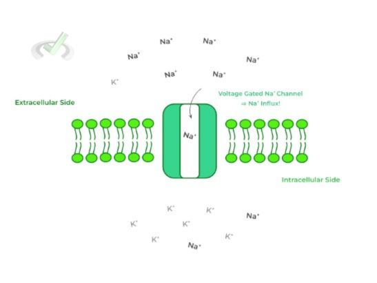
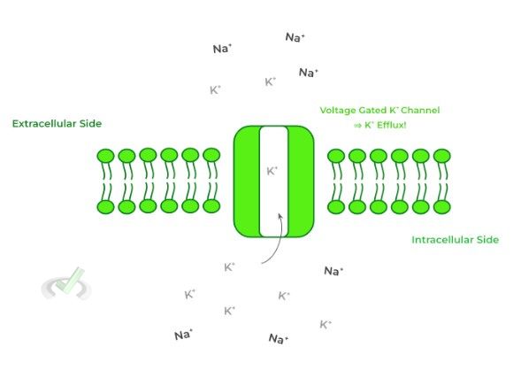
Additionally, another important aspect to note about the resting membrane potential is that Na⁺ ions are much concentrated/abundant in the EXTRACELLULAR space while K⁺ ions are more highly concentrated INTRACELLULARLY.
This will become very important when discussing the various influx and efflux of various ions in and out of the membrane throughout the action potential process.
II. Depolarization
After the threshold potential is reached, a process called depolarization occurs which results in making the membrane potential more positive to around a potential of +30 mV, as shown in the continued diagram below!
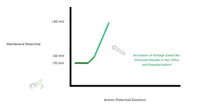
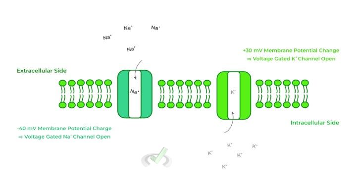
This results from the activation of what are called voltage gated Na⁺ ion channels — these ion channels are opened once the threshold potential is reached! Likewise, these result in the INFLUX of sodium ions down their concentration gradient because there’s a higher extracellular concentration of sodium ions than intracellularly!
III. Repolarization
After the membrane potential has reached +30 mV, this triggers the activation of another type of ion channel called voltage gated K⁺ channels which initiates the repolarization phase (i.e. making the membrane potential more negative).
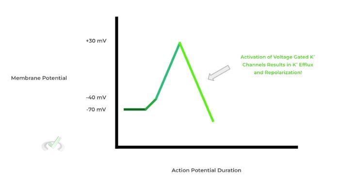
This occurs because of the EFFLUX of potassium ions out of the cell which causes the membrane potential to decrease because the K⁺ ions move down their concentration gradient since there’s less potassium ions extracellularly!
IV. Refractory Period/Hyperpolarization
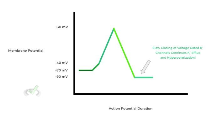
In this phase, the voltage gated K+ channels (which are slow to close compared to the Na+ channels) are still open and allowing for the efflux of K⁺ ions.
As a result, the membrane potential actually dips below the resting membrane potential and even goes down to about -90 mV.
From this point, we can talk about another important concept called refractory periods: simply put, these refer to time points during action potential propagation where another action potential CANNOT be initiated.
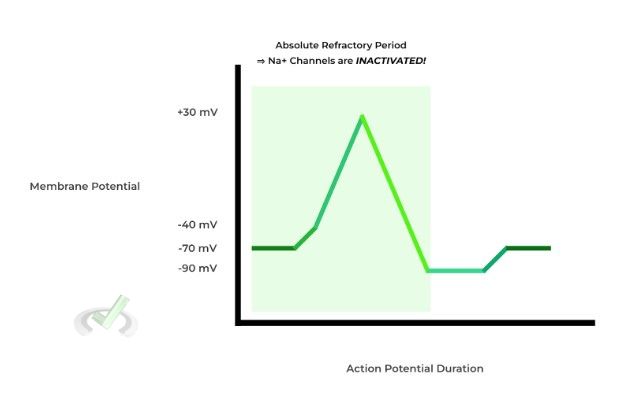
One type of refractory period is the absolute refractory period, which begins during depolarization and ends at the start of hyperpolarization During this period, another action potential CANNOT be fired no matter how strong the preceding stimulus is. This is because at this point the Na⁺ ion channels are inactivated!
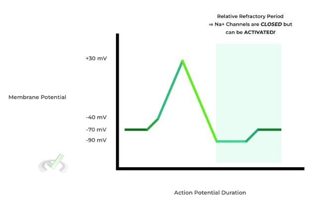
The relative refractory period occurs during hyperpolarization and is a rare case where another action potential can be generated, although the stimulus must be significantly stronger than that of a normal one.
In this case, another action potential can be generated because the Na⁺ channels are simply closed, but not inactivated per se. At this point, they’ve returned to their natural conformation and are waiting for another stimulus to open again.
To put it in an analogy (albeit, a little gross one), it’s similar to flushing a toilet: no matter how many times you press the lever, another flush can’t be initiated because the preceding flush is still making its way down the toilet bowl!
V. Returning to Resting Membrane Potential
After hyperpolarization, the membrane returns back to the resting membrane potential of -70 mV. This involves the return of the sodium and potassium ions to the proper location.
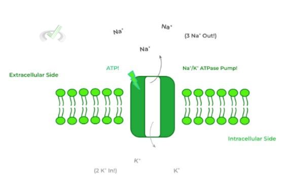
This is accomplished via a protein called sodium/potassium ATPase pump — this is an active transport protein which utilizes ATP in order to move 3 Na⁺ ions out of the cell and 2 K⁺ into the cell.
Take a look below at the complete diagram of the action potential!
III. Bridge/Overlap
We know there are a lot of different proteins to keep track of when learning about action potential generation and propagation. It definitely makes it easier to break them down into various categories to keep them a bit more organized.
I. Voltage Gated v.s. Ligand Gated Ion Channels
In our discussion of action potentials, it’s important to understand and categorize the different ion channels into either voltage gated or ligand gated ion channels!
Voltage gated ion channels are those that become activated and opened upon a change in membrane potential! These include the sodium and potassium gated ion channels which open in order to trigger depolarization and repolarization/hyperpolarization, respectively!
Conversely, ligand gated ion channels involve the binding of a ligand (i.e. signaling molecule ) in order to trigger the activation and opening of the ion channels. In this case, the postsynaptic dendritic receptors which bind to neurotransmitters are a great example of these receptors!
IV. Wrap Up/Key Terms
Let’s take this time to wrap up & concisely summarize what we covered above in the article!
A. Macroscale Propagation of the Action Potential
Whether an action potential will be initiated depends on the balance of excitatory and inhibitory neurotransmitters which bind to postsynaptic dendritic receptors. If the excitatory neurotransmitters predominate, an action potential is generated!
The action potential is generated at the level of the axon hillock and travels down the axon which sustains its electrical signal via the myelin sheaths.
When it reaches the axon terminal, it results in the activation and opening of Ca⁺² ion channels which allow for an influx of calcium — this triggers membrane fusion of vesicles with neurotransmitters in order to release them into the synaptic cleft!!
B. Action Potential Generation
Much of action potential generation is garnered by changes in membrane potential which in turn is governed via changes in ion influx and efflux!
I. Neurotransmitter Summation and Stimulus
In this stage, the binding of neurotransmitters to postsynaptic receptors will govern whether an action potential will be initiated.
Neurotransmitter binding needs to elicit a slight depolarization – from around -70 mV to -40 mV — in order to reach a threshold for an action potential to be generated!
II. Depolarization
If the depolarization threshold is reached, there’s a subsequent activation of voltage gated Na+ channels which allows for sodium influx into the cell and depolarization to a membrane potential of about +30 mV.
III. Repolarization
Once the membrane potential reaches +30 mV, the voltage gated Na+ channels become inactivated and close while voltage gated K⁺ channels open and allow for the efflux of potassium ions out of the cell!
IV. Hyperpolarization/Refractory Period
Because the voltage gated K+ channels are much slower to close compared to the voltage gated Na+ channels, hyperpolarization occurs where the membrane potential falls below its resting membrane potential.
V. Returning to Membrane Potential
Finally, in order to return the membrane potential back to its resting state, the Na⁺/K⁺ ATPase pump returns the ions to their respective location pumping 3 sodium ions out and taking in 2 potassium ions.
V. Practice
Take a look at these practice questions to see and solidify your understanding!
Sample Practice Question 1
A researcher discovers that a toxin from a plant inhibits the Ca⁺² ion channels located in the end of the axon terminal. Which of the following would be most directly affected if this toxin were to get into our system?
A. Insulation of Action Potential Propagation
B. Degradation of Neurotransmitters in Synaptic Cleft
C. Neurotransm itter Release
D. ATP Generation
Ans. C
Recall that its the influx of calcium ions at the axon terminal which triggers vesicle fusion in order to release the neurotransmitters contained within the vesicles into the synaptic cleft!
Sample Practice Question 2
ATP depletion would result in the dysfunction of which stage in action potential propagation?
A. Depolarization
B. Repolarization
C. Hyperpolarization
D. Return to Membrane Potential
Ans. D
This is a great example of how the MCAT would probably ask a question and test your knowledge via integrating multiple bits of information together.
Although we didn’t explicitly mention it in the article, the voltage gated sodium and potassium channels are all under passive transport since the ions are moving down their concentration gradient — recall also that passive transport does not require energy use in the form of ATP.
However, the sodium/potassium ATPase pump, which is active in order to return the neuron to its resting membrane potential requires ATP in order to actively transport these ions!






 To help you achieve your goal MCAT score, we take turns hosting these



















 reviews on TrustPilot