I. What is the Composition and Function of Blood?
Across the globe, transportation plays a huge role in getting people services, and good where they need to be. Likewise, we can think of the cardiovascular system as a similar transportation of the human body. It thanks to the cardiovascular system that nutrients, oxygen, and hormones are delivered throughout the body to keep all the organs working properly.
Though it may seem a little daunting at first with all the different arteries, veins and structures, it may be helpful to divide up the circulation into 2 different categories: 1) pulmonary circulation and 2) systemic circulation. If you can keep these 2 subdivisions in mind, it makes understanding the flow of the blood throughout the body.
II. Divisions of Blood Flow Throughout the Body
As mentioned above, we’ll focus on dividing the flow of blood throughout the body into pulmonary circulation and systemic circulation. We'll outline the flow of blood through a step by step process in order to make the flow of logic a little more simpler.
A. Pulmonary Circulation
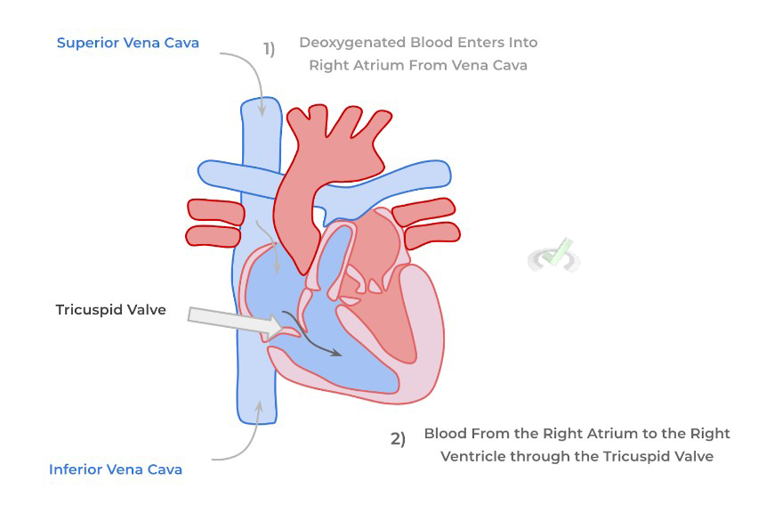
In pulmonary circulation, deoxygenated blood returning from systemic tissue is pumped towards the vasculature of the lungs in order to be reoxygenated. Recall that it’s the right side of the heart that receives deoxygenated blood coming from systemic tissue. ¹Deoxygenated blood within the body’s veins all converge into 2 main veins that feed into the right atrium: the superior and inferior vena cava.
²After the deoxygenated blood moves into the right atrium, it then moves into the right ventricle through the tricuspid valve. Recall that this valve is also termed the right atrioventricular valve given its location between the right atrium and ventricle.
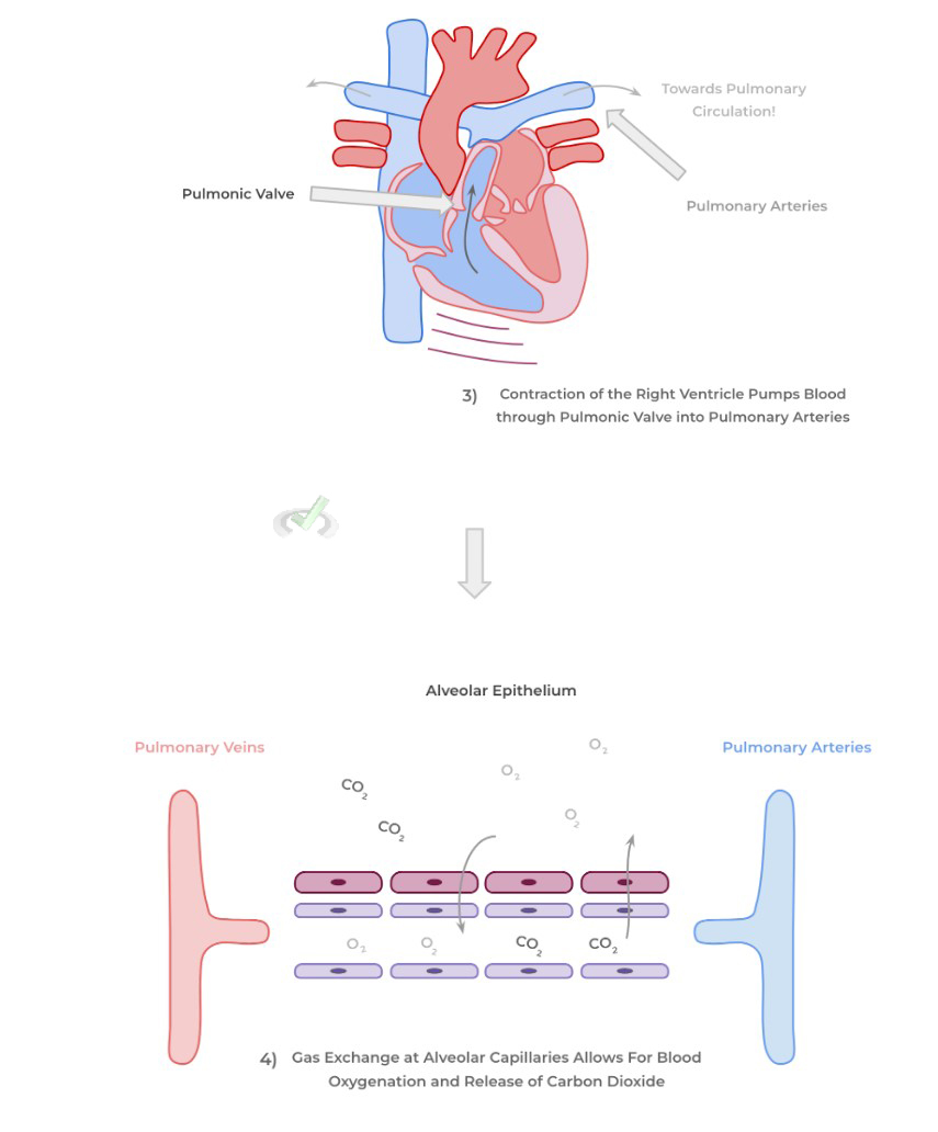
³Blood from the right ventricle is now pumped into pulmonary circulation via the contraction of the right ventricle! When the right ventricle contracts, it allows for the pumping of blood into the pulmonary arteries through the pulmonic valve (a semilunar valve).
If you noticed something a bit off, your intuition was right! Even though they are termed arteries (because they carry blood away from the heart), the pulmonary arteries are one exception where arteries actually are carrying deoxygenated blood (the other being the umbilical arteries)!
⁴The pulmonary arteries then transport the deoxygenated blood towards the alveolar capillaries where they can participate in gas exchange with the alveoli! Both the alveolar capillaries and epithelium are simple squamous epithelium to allow for efficient gas exchange!
B. Systemic Circulation
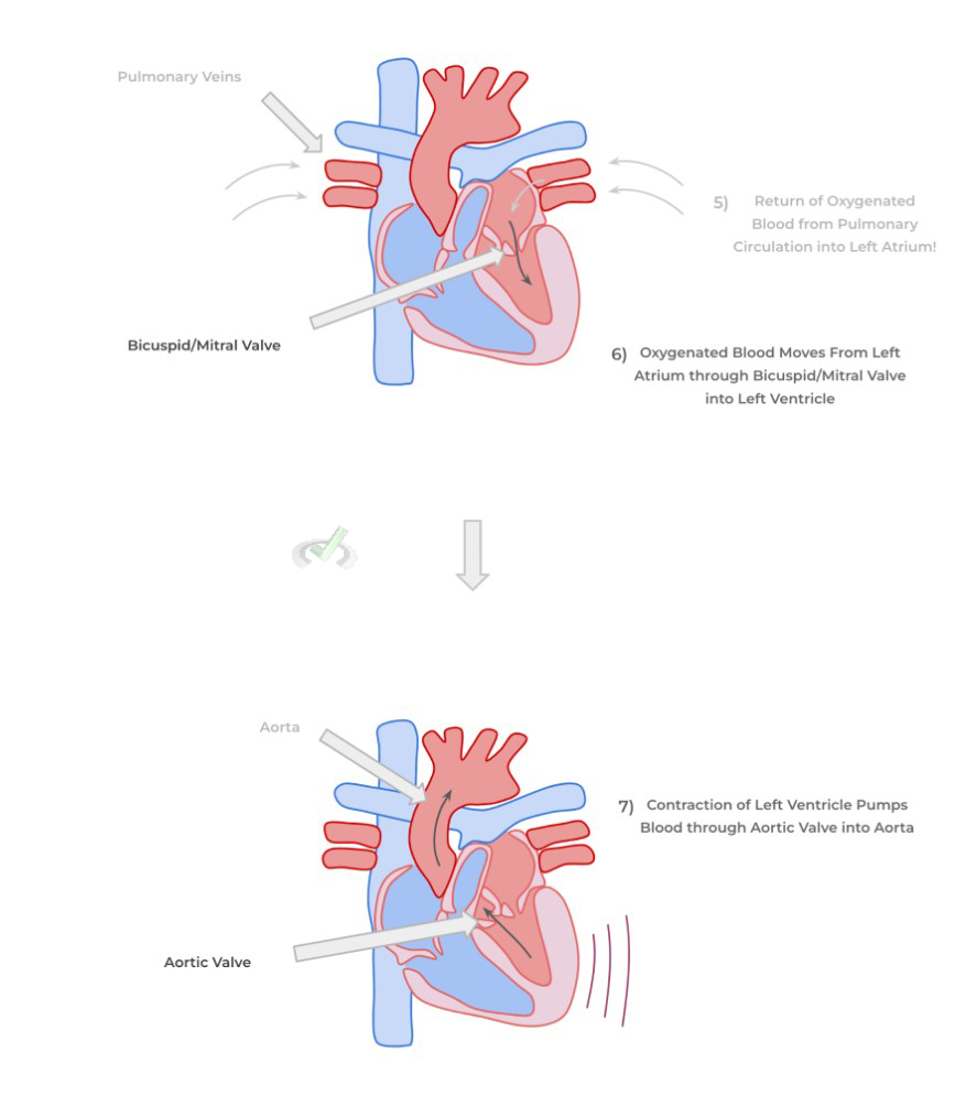
After blood has been reoxygenated in pulmonary circulation, the blood is now ready to reenter systemic circulation and deliver the oxygen and nutrients to systemic tissue! ⁵Oxygenated blood returns pulmonary circulation into the left atrium via the pulmonary arteries. Also recall that the left side of the heart receives oxygenated blood to be returned into systemic circulation.
If you felt your intuition kick in again, you’re 2 for 2! The pulmonary veins — termed veins because they carry blood towards the heart — are another exception where veins actually are carrying oxygenated blood (the other exception being the umbilical veins)!
⁶Similarly, blood from the left atrium passes through the bicuspid/mitral valve and is pumped into the left ventricle. ⁷Finally, the left ventricle similarly contracts and pumps oxygenated blood through the aortic valve into the aorta, the real first main artery. The aorta then divides into many different arteries which then delivers oxygen and nutrients throughout the body!
Then as mentioned in our “Structure of the Cardiovascular and Lymphatic System” study article, oxygenated blood is delivered via arteries and eventually drains into capillaries which allow for gas and nutrient exchange for the systemic tissues.
The deoxygenated blood from the capillaries is then drained into veins which return the blood back to the heart, thus repeating the cycle!
C. Basic of Cardiac Cycle
You definitely won’t be expected to understand the various complexities of the cardiac cycle and how to read an EKG (trust me, you’ll want to postpone that for a while….). However, it doesn’t hurt to at least be acquainted with the basics and how they relate to blood flow within the heart!
The cardiac cycle basically refers to the various contractile and relaxation states of the atria and ventricles that allows for the pumping of blood and filling of the cardiac chambers! It can be divided into 2 main states: systole and diastole.
I. Systole
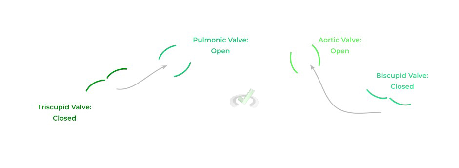
In its simplest form, systole refers to the portion of the cardiac cycle where both the right and left ventricle contract in order to pump blood into pulmonary and systemic circulation, respectively. As such, the pulmonic and aortic valve are open to allow for the flow of blood through their respective circulations.
Because of the high contractile pressures of the ventricles and the presence of the chordae tendineae and papillary muscles, the atrioventricular valves (tricuspid and bicuspid) close in order to prevent backflow of blood into the atria! This is actually what causes the first “lub” heart sound, termed S1.
II. Diastole
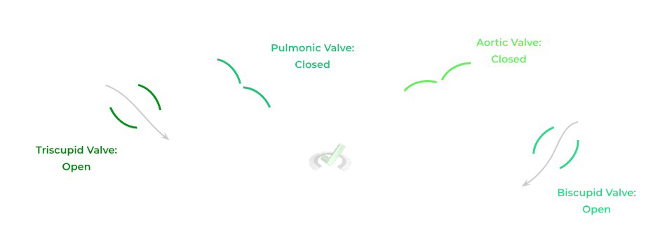
Conversely, diastole refers to when the ventricles relax in order to allow for the ventricles to be filled with blood coming in from the atria! As such, the tricuspid and bicuspid/mitral valve are open to allow for blood to flow in their respective cardiac chambers.
Because the ventricles are relaxing, there is no contractile force that’s generated: as such, the pulmonic and aortic valves are closed because no blood is being pumped into their respective circulations! The closure of these valves generates the second “dub” heart sound, termed S2.
III. Bridge/Overlap
A very cool aspect about the cardiovascular system is that you can apply some basics in regards to electric circuit design to understand how the various amounts of capillary networks don’t cause a high amount of resistance to blood flow.
I. Parallel Circuit Design of Capillary Network
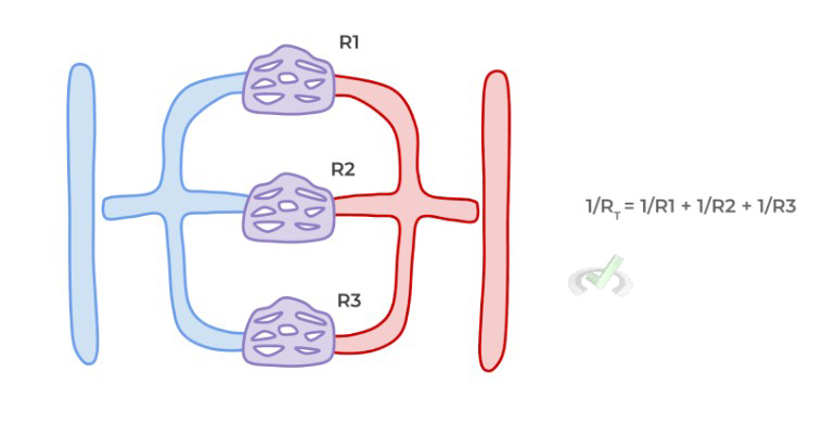
Essentially, each individual capillary network can be thought of as an individual resistance unit as seen in a basic electric circuit. Because there are so many capillary networks, the capillaries are arranged in parallel in order to minimize the amount of resistance encountered!
Recall when you place resistance units in parallel v.s. in series, the total summated resistance in parallel is significantly less than that of the in series pattern because you’re adding by the reciprocal, as shown below!
IV. Wrap Up/Key Terms
Let’s take this time to wrap up & concisely summarize what we covered above in the article!
A. Pulmonary Circulation
Deoxygenated blood first returns from systemic tissue from veins which drain into the main veins: the superior and inferior vena cava. These drain the blood into the right atrium which passes through the tricuspid valve to go into the right ventricle.
The right ventricle then contracts which pumps blood through the pulmonic valve into the pulmonary arteries which then takes this blood into the alveolar capillaries. It’s at the level of the alveolar capillaries that gas exchange can take place and blood can be reoxygenated.
B. Systemic Circulation
The reoxygenated blood is then taken back to the heart via the pulmonary veins which drain the deoxygenated blood to the left atrium. The blood then passes from the left atrium to the left ventricle via the bicuspid/mitral valve.
Finally, the left ventricle contracts which allows for the pumping of blood through the aortic valve into the aorta. The aorta then branches off into many arteries which branch into capillaries to allow for the delivery of oxygen and nutrients. Veins drain deoxygenated blood from the capillaries and the process repeats itself.
C. Basics of Cardiac Cycle
The cardiac cycle refers to the various contractile and relaxation states of the cardiac chambers which allows for the proper flow of blood within the heart! It contains 2 main phases: 1) systole and 2) diastole.
I. Systole
In this phase of the cardiac cycle, the ventricles are actively contracting which allows for the pumping of blood into pulmonary and systemic circulation.
As such, the pulmonic and aortic valves are open while the tricuspid and bicuspid/mitral valves are closed due to the high contractile pressure and presence of the chordae tendineae and papillary muscles.
II. Diastole
In contrast, diastole refers to the portion of the cardiac cycle where the ventricles are relaxing to allow for the ventricular filling of blood from the atria! As such, the tricuspid and bicuspid/mitral valves are open while the pulmonic and aortic valves are closed.
V. Practice
Take a look at these practice questions to see and solidify your understanding!
Sample Practice Question 1
In a pulmonary embolism, a blood clot is formed and usually gets lodged within pulmonary arteries.. As such, which of the following would experience an increase in blood flow?
I. Right Ventricle
II. Left Atrium
III. Left Ventricle
A. I only
B. II only
C. I and II
D. II and III
Answer: A. I only
If a clot becomes lodged at the level of the pulmonary artery, this results in blood not being able to move blood through pulmonary circulation to reach systemic circulation. However, the clot will result in a backflow of blood into the right side of the heart because the blood within the pulmonary arteries originated from the right side of the heart.
Sample Practice Question 2
Complete the following statement.
Contraction of the __________ ventricle results in opening of the ___________ valve and closure of the __________ valve during ___________
A. Left, Pulmonic, Bicuspid, Systole
B. Right, Pulmonic, Tricuspid, Systole
C. Right, Pulmonic, Tricuspid, Diastole
D. Left, Aortic, Bicuspid, Diastole
Answer: B. Right, Pulmonic, Tricuspid, and Systole
This answer is the only combination of terms which correctly fits the sequence of right sided heart contraction during systole.







 To help you achieve your goal MCAT score, we take turns hosting these





















 reviews on TrustPilot