I. What is the Anatomy of the Digestive System?
The digestive system is one of the most intricate and elaborate body systems. It comprises multiple organs and types of tissues, which all work together to break down food into vital substrates and nutrients, which are then used for energy throughout the body.
Today, we’ll examine the digestive system's various organs and how their main and unique functions contribute to the overall digestive process. This process involves ingesting food, its breakdown in the stomach and small intestine, absorption of nutrients in the small intestine, and the elimination of waste. Let’s get started!
II. The Organs of the Digestive System
The digestive system's organs can be divided into those comprising the alimentary canal (a.k.a. the gastrointestinal tract) and accessory organs. The alimentary canal refers to the organs that form a continuous pathway for food from ingestion at the mouth to its eventual defecation at the rectum.
Accessory organs, such as the liver and gallbladder, are not contained within the alimentary canal but still play an active role in digestion through other processes.A. Principal Organs of the Alimentary Canal
Again, to reiterate, the organs of the gastrointestinal tract are continuous, like a tube or a highway. Each organ has a specific function, which ultimately helps the food digest and progress towards the end of the canal.
I. Esophagus
The esophagus essentially serves as a connection between the oropharynx and the stomach and propels the food bolus down towards the stomach via peristalsis, the rhythmic contraction of the surrounding smooth muscle.
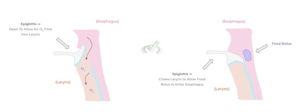
The other main high-yield point is a vital structure composed of elastic cartilage called the epiglottis, which resembles a flap.
Because the oropharynx has openings towards the esophagus and respiratory tract, the epiglottis ensures that while we swallow, none of the contents will enter the windpipe and cause obstruction by blocking the opening to the windpipe, specifically the larynx.
II. Stomach
Though the oral cavity initiates digestion via mastication and other digestive enzymes (e.g., salivary amylase), most digestion begins in the stomach.
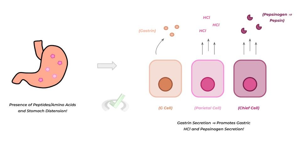
This is due to the highly acidic nature of the stomach lumen, in addition to the various hormones and enzymes released, which promote digestion. Some important gastric cells to know regarding what substrate they release and their function include the following:- G Cells: Secretes gastrin hormone ⇒ Promotes release of HCl acid by parietal cells and pepsinogen release by chief cells
- Chief Cells: Secrete pepsinogen (active form: pepsin) ⇒ Main gastric proteolytic enzyme
- Parietal Cells: Secrete HCl ⇒ This acidic substance is present in the stomach and promotes digestion. It also activates pepsinogen to the active pepsin form.
III. Small Intestine
Following the stomach is the small intestine, which can be divided into three sequential parts: the duodenum, the jejunum, and the ileum. In addition to continuing digestion with various enzymes, the small intestine is where the majority of absorption takes place.
Its primary role in absorption is due to various unique characteristics in its cellular architecture and various transporter proteins. The epithelium of the small intestine is unique as it contains villi, which are “projections” and folds of the small intestine epithelium that work to increase the surface area of digestion!
The surface area is further increased as the individual cells of the small intestine epithelium contain microvilli, extensions and projections of the cells! To recap, villi are projections of the SI epithelium, while microvilli are projections of the individual cells.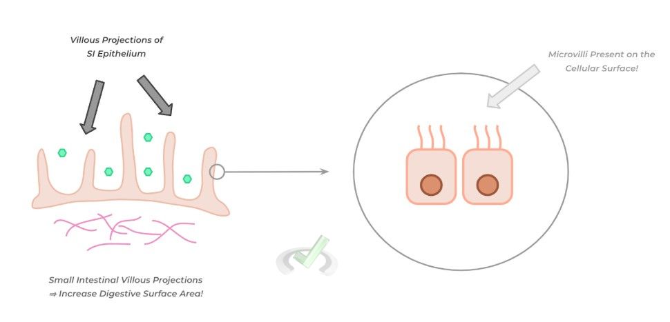
As mentioned above, the small intestine is the primary organ for the body's absorption of necessary nutrients and water. Absorption mostly occurs in the duodenum, with the jejunum and ileum absorbing remaining substrates and nutrients.
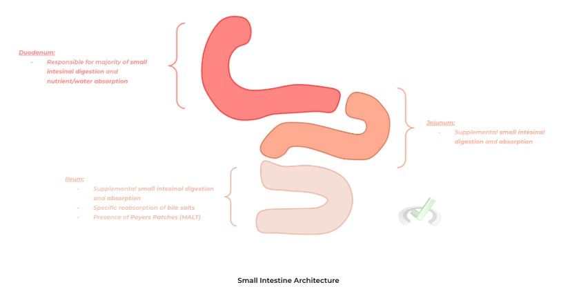
However, the ileum has some distinct characteristics that are not found in the other portions of the small intestine. For instance, only the ileum is responsible for the reabsorption of bile salts, which are released by the gallbladder and are essential for digesting incoming fats.
Additionally, underneath the ileal epithelium is mucosal-associated lymphoid tissue called Peyer's patches—this tissue resembles that found in lymph nodes, where abundant lymphocytes are ready to proliferate in the case of a GI infection!IV. Large Intestine
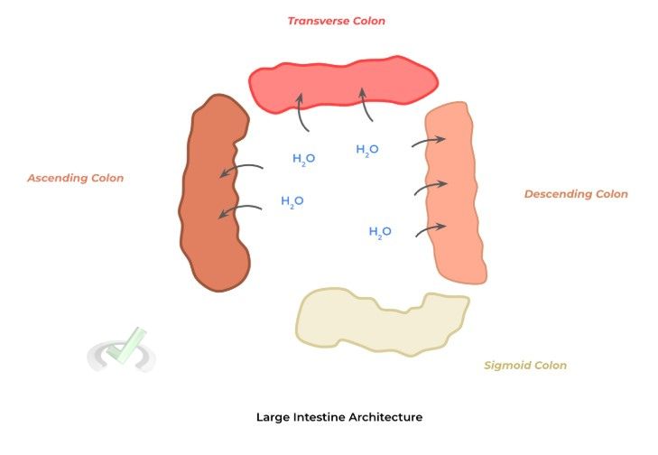
Finally, the large intestine follows the small intestine, which can similarly be divided into sequential parts: the ascending colon, the transverse colon, the descending colon, and the sigmoid colon.
The small intestine's sole function is the absorption of water in addition to water-soluble vitamins (i.e., vitamin B and C).
However, it’s important to note that most water reabsorption occurs in the small intestine — the large intestine simply absorbs the residual water. Still, it does not absorb other substances like glucose or amino acids.
B. Accessory Gastrointestinal Organs
As mentioned above, these gastrointestinal organs are not continuous with the alimentary canal but still function in the digestive process, often secreting substances that aid in digestion. These include the liver, gallbladder, and pancreas.
I. Liver and Gallbladder
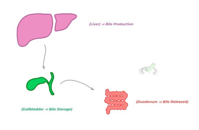
Being our body’s largest internal organ, the liver has a multitude of functions necessary for our body to function. However, in the context of gastrointestinal function, the main function of the liver is to produce bile.
We will discuss bile's function in another article, but in summary, it aids in the digestion of fatty substances via a process called emulsification. After being produced in the liver, bile is stored in the gallbladder.
When food enters the small intestine, it triggers the contraction of the gallbladder, allowing the release of the stored bile into the duodenum to further aid in fat digestion.
II. Pancreas
Finally, the other accessory gastrointestinal organ is the pancreas. Again, we cover the primary digestive function of the pancreas in another article — for now, understand that the pancreas stores and releases pancreatic fluid.
Various digestive enzymes, including proteases, lipases, and amylases, are present. Similar to the gallbladder, the pancreas releases pancreatic fluid into the duodenum when food enters the small intestine to further aid digestion.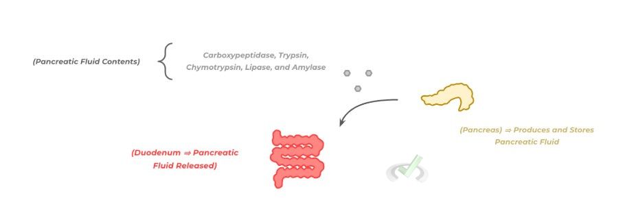
III. Bridge/Overlap
While our first inclination is to associate the liver and pancreas as gastrointestinal organs because of their proximity to the GI tract, it’s also important to note that they have other functions related to different parts of the human body. Let’s quickly review them!
I. Non-Gastrointestinal Functions of the Liver and Pancreas
The liver is essential in producing bile and oncotic proteins, particularly albumin. Oncotic proteins help maintain the oncotic pressure within blood vessels in order to maintain volume and blood pressure homeostasis.
Other liver proteins include angiotensinogen and various acute-phase reactant proteins, which are innate immune proteins produced in response to infection.
Finally, the liver plays a major role in detoxifying various drugs and metabolites, specifically via a group of enzymes collectively known as the cytochrome P450 system.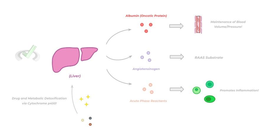
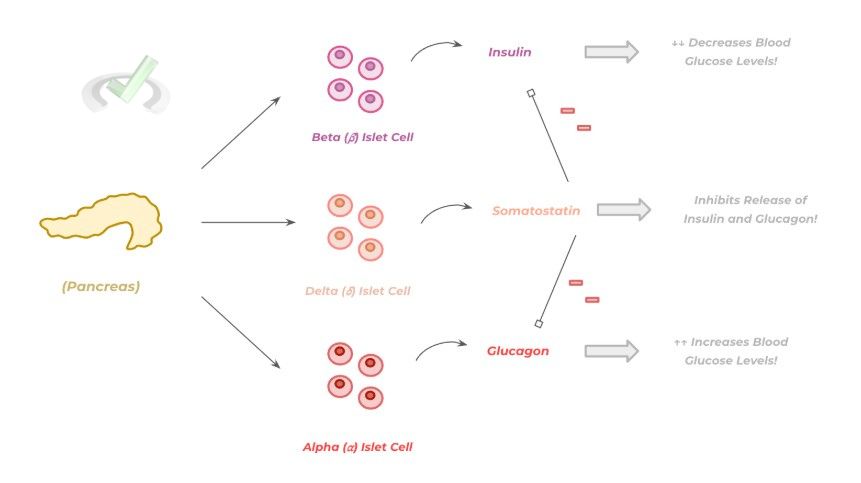
Additionally, recall that the pancreas is an endocrine organ, particularly in regulating glucose homeostasis. Look at the figure below for a quick review of the hormones the pancreas produces and their functions!
IV. Wrap Up/Key Terms
Let’s take this time to wrap up & concisely summarize what we covered above in the article!
A. Principal Organs of the Alimentary Canal
The main organs of the alimentary canal include the esophagus, the stomach, the small intestine, and the large intestine, which together form a continuous digestive canal where food can be digested.
I. Esophagus
After food begins digestion in the oral cavity, the food bolus enters the esophagus which propels the movement of the food bolus via peristalsis.
Because the oropharynx has openings towards the alimentary canal and the respiratory system, a specialized piece of elastic cartilage called the epiglottis, which covers the larynx during swallowing, prevents the food bolus from entering the respiratory tract.II. Stomach
Following the esophagus is the stomach, a J-shaped organ that has a massive role in digestion due to its highly acidic nature within the lumen and the presence of proteolytic enzymes.
Some of the important cells of the stomach include the G cells (which release gastrin hormone), chief cells (which release pepsinogen ⇒ pepsin), and parietal cells (which release HCl).
III. Small Intestine
The small intestine can be divided into three other segments: the duodenum, the jejunum, and the ileum. In addition to further digestion, the small intestine is where most nutrient and water absorption occurs, with a good portion occurring at the duodenum.
The ileum has particularly unique functions, where bile salt reabsorption occurs. In addition, the ileum is also a type of mucosal-associated lymphoid tissue (MALT) called Peyer’s patches, with growing lymphoid follicles!IV. Large Intestine
Like the small intestine, the large intestine is also segmented into the ascending colon, the transverse colon, the descending colon, and the sigmoid colon.
The main role of the large intestine is the absorption of water and water-soluble vitamins (e.g., vitamins B and C), though most of the water absorption is done at the level of the small intestine.B. Accessory Gastrointestinal Organs
Though not continuous with the alimentary canal, accessory gastrointestinal organs also aid in digestion via the production and secretion of substances to aid in this process.
I. Liver and Gallbladder
Though the liver has many functions, including producing various proteins and detoxifying, its main role as a gastrointestinal organ is to produce bile, which is then stored within the gallbladder.
As covered in another article, bile is essential for the reabsorption of fatty particles via emulsification. When food enters the small intestine, the gallbladder contracts, allowing for the bile to be released into the duodenumII. Pancreas
As an exocrine and gastrointestinal organ, the pancreas produces pancreatic fluid, which is rich in digestive enzymes, including various proteases, lipases, and amylases.
Similarly, when food enters the small intestine, the pancreas releases pancreatic fluid into the duodenal lumen to further aid digestion.
V. Practice
Take a look at these practice questions to see and solidify your understanding!
Sample Practice Question 1
A 34-year-old woman comes into the clinic complaining of increased heartburn, especially after she eats. After further history taking and a physical exam, a diagnosis of gastroesophageal reflux disease (GERD) is put high on the differential, and she is started on the medication pantoprazole, which works to decrease the acidity in her stomach. Which of the following cells is most likely inhibited by this drug?
A. G cells
B. Parietal Cells
C. Chief Cells
D. Delta Cells
Ans. B
Parietal cells are most likely inhibited because recall that the parietal cells release HCl, creating the stomach lumen's highly acidic environment.
Specifically, pantoprazole is a proton pump inhibitor that decreases stomach acidity, which is a possible treatment for GERD.
Sample Practice Question 2
A 45-year-old man who came to the clinic for chronic abdominal pain underwent a colonoscopy, which is significant for the presence of a possibly malignant polyp within the ileum because the polyp has already grown past the submucosal layer; therefore, a resection (removal) of the ileum is recommended, which comes with physiological consequences. Which of the following would the patient most likely suffer from following the surgery?
A. Increased presence of fatty stool
B. Increased absorption of fats
C. Decreased absorption of water
D. Increased absorption of vitamin A, D, E, and K
Ans. A
This is another good way the MCAT might want to test you on your content knowledge and application. Because the ileum is responsible for the reabsorption of bile salts, this process would be hindered in a case of a partial removal of the ileum.
Because bile is essential for absorbing fatty substances (and fat-soluble vitamins), decreased bile absorption would lead to reduced absorption of fat (and fat-soluble vitamins), increasing fatty stools.




 To help you achieve your goal MCAT score, we take turns hosting these


 reviews on TrustPilot