I. What are the Various Processes of Embryogenesis?
We know…. looking at the title of this article can be really daunting (we’ve definitely been there before!). However, despite it being scary superficially, the study of embryology is truly fascinating when you think about how one cell can eventually develop into a whole person!
When approaching the study of embryology, we’ll definitely try our best to give you the basic foundation and really emphasize topics that the MCAT will likely test you on — they’re not looking at you to become a master embryologist but to simply understand the basics and apply them to other MCAT topics.
II. From Zygote to Person
As amazing as it sounds, it’s truly incredible how one single cell can eventually develop into the 30 trillion cells that make up your entire body. Let’s go ahead and outline the basic steps that take place in order for this process to occur!
A. Zygote Formation, Cleavage, and Blastulation
The entire process of embryogenesis begins with fertilization of the 2 haploid gametes (a sperm and egg cell) eventually resulting in a diploid, zygote cell!
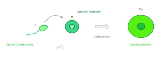
From here, the zygote undergoes a series of rapid cell divisions known as cleavage. It’s important to note that during these cell divisions, the cells are genetically identical but become smaller in size, as they lose cytoplasm.
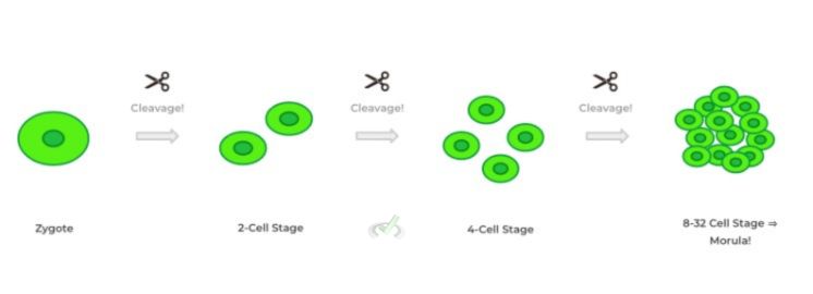
These cell divisions not only generate the cells needed for embryogenesis but also adjust the nucleus to cytoplasm ratio! When about 8-32 cells are present after cleavage, the resulting structure is called a morula (Latin for “mulberry”).
The morula then makes its way towards the uterus and begins the process of blastulation in which a structure called the blastocyst is formed — the key word to associate with this process is “differentiation”, as they’re well be multiple new cell types produced!
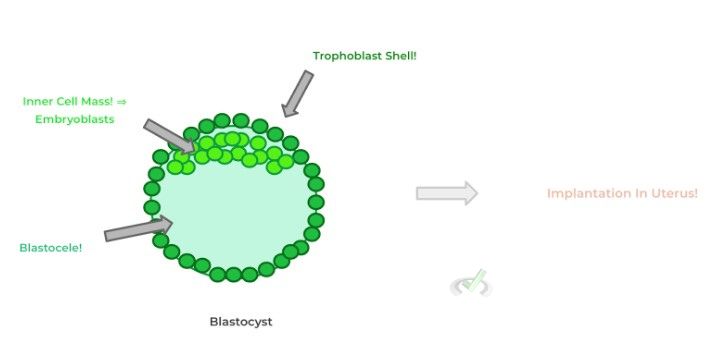
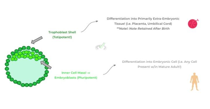
As shown above, the cells of the morula proliferate, migrate, and differentiate into 2 new cell types: the trophoblasts and the embryoblasts. The trophoblasts will form the outer shell of the blastocyst while the embryoblasts remain inside the blastocyst and clump together in a complex called the inner cell mass!
The trophoblasts are special in the sense that they’ll differentiate into cells which are crucial for the support of the embryo such as the placenta and umbilical cord! Conversely, the embryoblasts of the inner cell mass have the potential to give rise to any of the cells present within the adult human body!
B. Gastrulation
After the formation of the blastocyst, a process called gastrulation occurs which forms a 3 layered structure called the gastrula.
While the trophoblast differentiate into the cells which support embryo growth, the embryoblast of the inner cell mass begin to further differentiate and migrate to form 3 cell layers: the ectoderm, mesoderm, and endoderm.
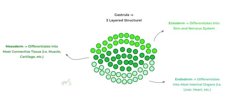
The cells located in each of these 3 layers are multipotent with each layer of cells being committed to differentiate into specific tissue types.
The ectoderm most notably differentiates into the skin and nervous system while the mesoderm differentiates into most connective tissue (i.e. cartilage, bone, etc.) as well as muscle tissue! Finally, the endoderm cells will primarily differentiate into most of the body’s internal organs such as the heart, lung, liver, etc.
C. Neurulation
The last main part of embryogenesis that the MCAT will likely cover is the basics of neurulation, the development of the nervous system — we know…. It may seem a little scary at first but we’ll make sure to lay out the process step by step and give you only the basic information needed!
The first thing to note is that a portion of the mesoderm develops a structure called the notochord, which is basically a specialized, differentiated portion of the mesoderm.
The notochord then sends molecular signals to the overlying ectoderm which causes a portion of the ectoderm to thicken into a structure called the neural plate!
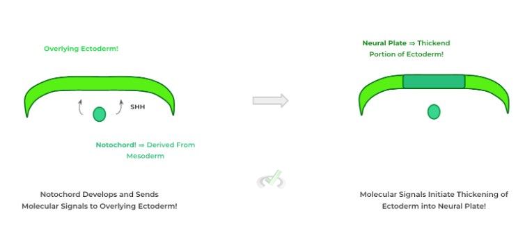
Following its formation, the neural plate begins to invaginate inwardly resulting in the formation of a neural groove. At the top part of the invaginations are neural folds which will come close together and eventually fuse!
The combined fusion and invagination creates a structure called the neural tube which will eventually develop into the main structures of the CNS including the brain and spinal cord!
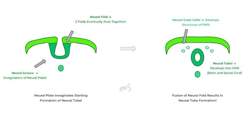
Another structure, not explicitly shown during the neural tube formation are the neural crest cells — these cells also emerge from the neural folds and reside on the periphery of the neural tube and will develop into structures of the peripheral nervous system!
III. Bridge/Overlap
During our discussion about blastulation and gastrulation, we talked about how the cells eventually proliferate and differentiate into cells such as the trophoblasts and embryoblasts — it’s important to note that these cells can give rise to different cell types depending on their potency as we’ll discuss!
I. Totipotent v.s. Pluripotent v.s. Multipotent
All these 3 terms can be used to distinguish the differentiation potential of these different stem cells — as such, these terms can also be used to describe the different cells of the blastocyst and gastrula as they differ in what type of cells that can develop into!
Trophoblasts have a totipotent meaning as they have the potential to differentiate into extraembryonic and embryonic cells! Extra-embryonic cells are cells which form the structures which help support embryonic growth and are not retained in adult life such as the placenta and umbilical cord.
Conversely, neuroblasts are pluripotent meaning they have the ability to develop into any embryonic cell which is basically any cell that’s present in a mature adult.
Finally, the cells of different gastrula layers (i.e. ectoderm, mesoderm, and endoderm) are best termed multipotent as they are more limited in the cells they can differentiate into. Think of how only ectodermal, multipotent cells give rise to the cells of the skin and CNS!
IV. Wrap Up/Key Terms
Let’s take this time to wrap up & concisely summarize what we covered above in the article!
A. Zygote Formation, Cleavage, and Blastulation
A diploid zygote is formed upon the fertilization of a haploid egg with a haploid sperm cell. From here, the zygote undergoes a series of cellular replication called cleavage in order to produce more cells and adjust the nucleus to cytoplasm ratio.
When the cleavage process produces about 8-32 cells, the structure is termed the morula. From here the morula can then begin the process of blastulation to form the blastocyst.
The blastocyst is a spherical structure containing a trophoblastic shell and embryoblasts within the structure bundled together in a complex called the inner cell mass.
Trophoblasts primarily will differentiate into extraembryonic cells (those which primarily support embryonic growth) while embryoblasts will develop into embryonic cells, which is basically any cell present within an adult human body!
B. Gastrulation
After implantation and formation of the blastocyst, gastrulation occurs which now forms 3 main cell layers: the ectoderm, mesoderm, and endoderm which arise from the embryoblasts in the inner cell mass!
The ectoderm will primarily differentiate into the skin and nervous system structures while the mesoderm develops into much of the main connective tissue such as bone, cartilage and other tissue such as muscle! The endoderm will primarily differentiate into much of the body’s internal organs!
C. Neurulation
The preliminary development of structures which eventually give rise to the structures of the nervous system is called neurulation. First, a differentiated portion of the mesoderm called the notochord sends molecular signals to the overlying ectoderm to cause their thickening into a neural plate.
The neural plate then invaginates and forms a neural groove with overlying neural folds. Eventually the folds fuse together closing the groove and forming the neural tube which will eventually develop into the CNS! Neural crest cells also emerge and will develop into the structures of the PNS!
V. Practice
Take a look at these practice questions to see and solidify your understanding!
Sample Practice Question 1
An embryologist is examining a developing embryo and identifies a group of cells which differentiate and form cells for the chorionic villi, an important structure of the placenta. At which step is the earliest point you can identify these cells that will differentiate to form the chorionic villi?
A. Fertilization
B. Blastulation
C. Cleavage
D. Neurulation
Ans. B
In blastulation, a structure called the blastocyst forms which consists of a trophoblastic shell and an inner cell mass composed of embryoblasts.
The trophoblasts are special as they are totipotent meaning they can differentiate into both extra-embryonic and embryonic structures. Extra-embryonic structures are those that support the growth of the embryo and are not retained in adulthood such as the placenta. As such, since trophoblast emerges during blastulation, this is the earliest point at which we can identify the cells.
Sample Practice Question 2
Spina bifida is a congenital condition in which there’s a malformation of the spinal cord possibly causing paralysis. Which of the following structures might have had an unsuccessful formation and which germ cell layer does it derive from?
A. Neural Crest Cells, Ectoderm
B. Neural Tube, Ectoderm
C. Notochord, Ectoderm
D. Neural Tube, Mesoderm
Ans. B
Recall that after fusion of the neural folds, the neural tube (which is derived from the ectoderm from the neural plate) will develop into the main structures of the CNS such as the brain and spinal cord.
Being that the spinal cord is not developed properly, there may be a problem with the maturation or development of the neural tube. Recall also that the neural crest cells are responsible for the development of the PNS.
Note also that the notochord could have also had a problem; however, recall that the notochord is derived from the mesoderm, not the ectoderm as stated in choice C.







 To help you achieve your goal MCAT score, we take turns hosting these