I. How is the Integumentary System Structured?
With so many avenues for its promotion, from advertisements, social media, etc., skin care has taken the world by storm! From Youtube dermatologists giving advice to beauty empires like Sephora and Olive Young, there’s definitely been a lot more focus recently on how we can keep our skin looking nice and healthy!
But considering all the products and procedures we put on our skin, it definitely begs the question, “What is our skin made of and how is it structured?”. We hope after reading this article, we can give you guys a little more insight on this question. Let’s get started!
II. Divisions of the Integumentary System
Going back to its Latin root, the name for the integumentary system actually comes from the Latin word integumentum which means “a covering” — the Latin meaning is so useful in describing the main function of this system as it acts as a covering to shield the external world from our internal organs!
A. Skin
As you may have heard, the skin is the largest overall organ of the human body, not to be confused with the largest internal organ which is the liver! There are 3 main layers of the skin all composed of different tissue and cell types: the epidermis, dermis, and hypodermis/subcutaneous tissue.
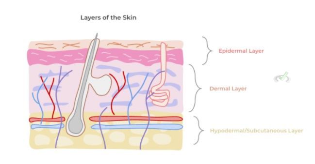
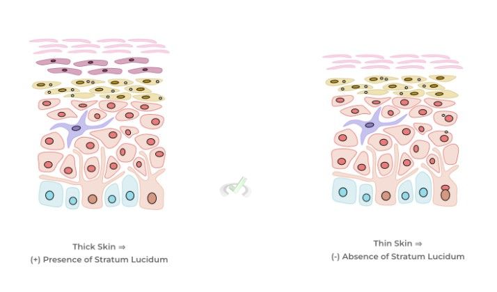
It’s important to note that there actually are 2 types of skin tissue within the body: thick and thin skin! As indicated by the name, thick skin has a much more substantial layer of cells than thin skin due to an extra layer of cells within the epidermis which we’ll get into!
Thick skin is found ONLY at the palm and soles of the hand and feet as these added cell layers help support these sites because of the great amount of external force the experience on a day to day basis. Conversely, thin skin is found throughout all the other parts of the body.
They also differ in the structures they house as thin skin houses hair follicles, sebaceous (oil producing glands) and sweat glands while thick skin ONLY houses sweat glands. This is easy to think about if you remember that your palms and sole don’t usually have hair or oil associated with them!
I. Epidermis
This layer is the most superficial layer of the skin and is the one most complex as it contains many cell layers! Look at the diagram below for a complete view of the layers and we’ll break them down a couple of layers at a time!
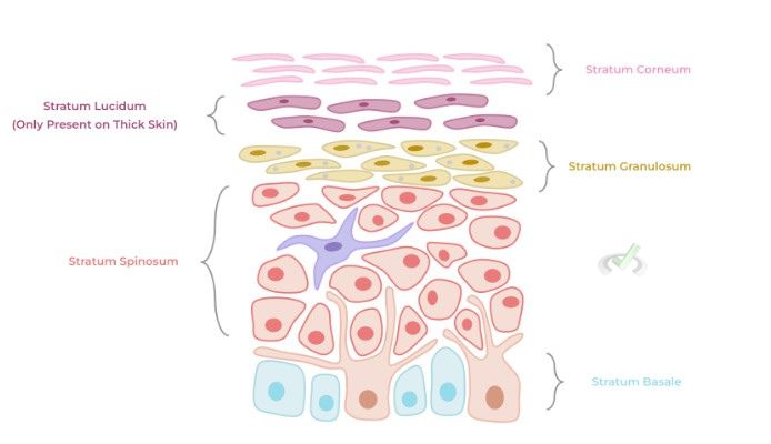
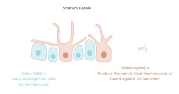
The stratum basale is the bottom most layer of the epidermis and houses the stem cells which serve as progenitors to the cells (called keratinocytes) located above the layer.
The stratum basale also houses melanocytes which produce the pigment, melanin. This pigment is transported to the superficial epidermal layers to coat the keratinocytes as a means to protect against the harmful UV radiation!
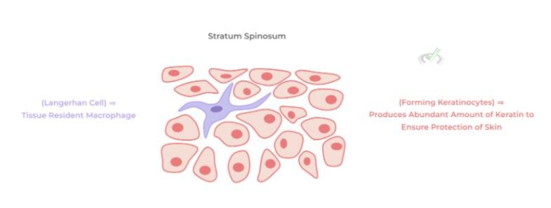
The stratum spinosum is the next layer and is the layer where keratinocytes begin to form — these are the main cells of the epidermis as they contain an abundant amount of keratin, which is a fibrous protein important in regards to skin protection!
This layer also houses the tissue resident macrophages called Langerhan cells which allow for the phagocytosis and presentation of foreign antigens!
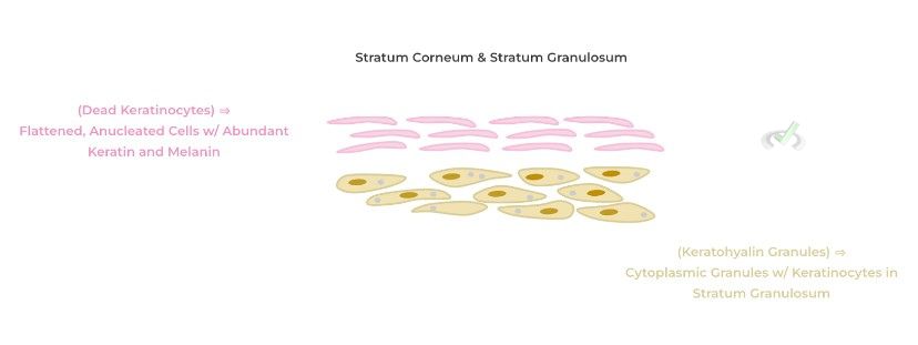
The stratum granulosum gets its name from the presence of keratohyalin granules within the keratinocyte cytoplasm.
In addition to keratin, these granules have other proteins which aid in the process of degrading the various organelles of the cells such as the nucleus, mitochondria, etc. as well as adopting a more flattened cell shape!
After all the organelles are removed and the cell adopts this flattened confirmation, they’re pushed into the last layer called the stratum corneum. All that remains within these dead cells are an abundant amount of keratin protein and melanin — these are in fact the dead cells which are shed from the skin in a process called desquamation!
Finally, the last remaining layer to discuss is the stratum lucidum — the most important thing to know about this layer is that it’s only found in THICK SKIN which if you can recall is the skin present on the palm and soles!
II. Dermis
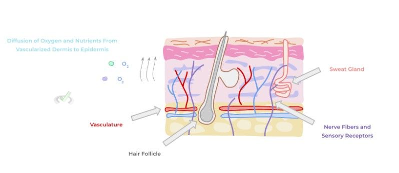
Below the epidermis is the dermis which is primarily composed of dense connective tissue and is connected to the epidermis via the basement membrane. The most important concept to understand about the dermis is that it’s vascularized and innervated!
The dermis contains an abundant amount of blood vessels, nerves and sensory receptors, and also houses other structures such as sweat glands and hair follicles! In contrast, the epidermis is avascular as it lacks any blood vessels — as a result, nutrient and oxygen supply to the epidermis occurs via diffusion from the dermal vasculature!
II. Hypodermis/Subcutaneous Layer
Finally, the bottom most layer of the skin is the hypodermis/subcutaneous layer which is primarily composed of fat cells, also called adipocytes.
The abundant amount of fat allows for this layer to function as a cushioning for our internal structures if there’s external trauma as well as functioning as an insulator in order to retain heat!
B. Glands
As mentioned before, there are various secretory glands spread out throughout the body located on our skin, specifically the dermis! There are 2 main types of glands: sweat glands and sebaceous glands — let’s go ahead and take a look at them a little more!
I. Sweat Glands
As their name implies, these secretory glands are responsible for producing sweat which is essentially water rich in sodium that’s released as a means for cooling down the body. There are 2 main types of sweat glands as well: eccrine and apocrine sweat glands.
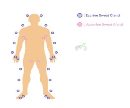
Eccrine sweat glands are found all throughout the body and are particularly abundant in the palms and soles! Apocrine sweat glands are more localized in areas of the body active in puberty such as the armpits, groin, nipples, etc.
II. Sebaceous Glands
Also located in the dermis, sebaceous glands are glands which produce and secrete oil in order to provide a lipophilic barrier to prevent water loss from the skin!
Usually, sebaceous glands are associated and secrete oil into hair follicles as we’ll touch on in a bit. Additionally, recall that hair follicles and sebaceous glands ARE NOT present in thick skin!
C. Hair and Nails
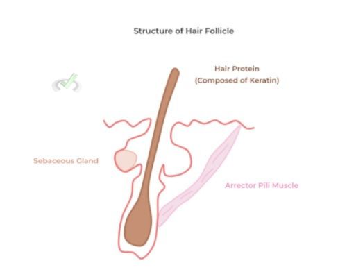
Note also that the hair and nails are composed of the same fibrous keratin protein of the skin’s keratinocytes. The hair follicle, again localized in the dermis, is essentially a specialized portion of the skin which contains a tubular shaft to allow for the growth and housing of the hair.
Associated with the hair follicle are sebaceous glands which again release oil to both provide a hydrophobic barrier to the skin and nourish the growing hair! In addition, the hair follicle also has an associated smooth muscle called an arrector pili muscle which can contract and lift the hair and can be utilized as a means of heat retention!
III. Bridge/Overlap
You might look and study some of the basic biochemical concepts and wonder “how will this really be applied to the human body?”. We totally get it — we were in your same shoes before! But in this bridge section, we’ll give you an example of how our hair is actually an example of a secondary structure of protein!
I. Keratin Secondary (2˚) Structure in Hair
Recall that after the 1˚ protein structure — which is simply the linear amino acid sequence —- is the 2˚ structure which refers to how the hydrogen bonds between the carbonyl and amino groups interact with each other.
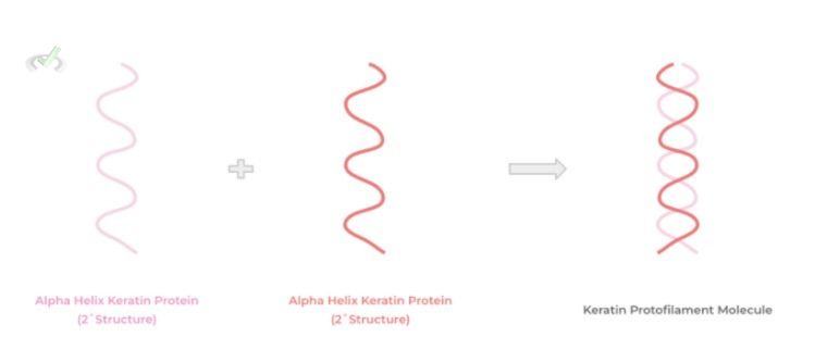
There are 2 possible secondary structures that can be formed: an alpha helix chain and a beta-pleated sheet. The keratin proteins of hair are actually arranged in an alpha helix structure! When 2 of these keratin alpha helices come together, they form a protofilament keratin molecule!
IV. Wrap Up/Key Terms
Let’s take this time to wrap up & concisely summarize what we covered above in the article!
A. Skin
The skin is composed of 3 main layers: the epidermis, dermis, and the hypodermis/subcutaneous layer — each of these layers has different compositions of cells and tissue, all of which have a distinct function!
Skin can be divided into thick and thin skin. Thick skin has an extra cell layer located in the epidermis and is primarily found in the palms and soles of the hands and feet — this type of skin has sweat glands but lacks any hair follicles or sebaceous glands. Thin skin is found throughout the rest of the body and contains all types of glands and hair follicles!
I. Epidermis
The stratum basale contains the stem cells which serve as progenitors to keratinocytes as well as melanocytes which produce melanin to protect against UV radiation.
The stratum spinosum is the layer where the keratinocytes begin to form as they begin to produce keratin, a fibrous protein for skin protection. In addition, this layer also holds the Langerhan cells which are the tissue resident macrophages.
The stratum granulosum is known for the presence of keratohyalin granules which aid in the process of removing the keratinocytes’ organelles and having them adopt a flattened shape.
Finally, the stratum corneum contains all the dead keratinocytes which only contain keratin protein and the melanin pigment. Note that the stratum lucidum is only found in thick skin!
II. Dermis
Below the epidermis is the dermis, which are connected to one another via the basement membrane! The dermis is primarily composed of connective tissue and is highly vascularized and innervated while also housing various glands and hair follicles.
Because the corresponding epidermis is avascular, it relies on diffusion of oxygen and nutrients from the dermis in order to be properly nourished!
III. Hypodermis/Subcutaneous Layer
This layer is composed entirely of fat cells (adipocytes) and serves as a role in cushioning of the internal organs as well as heat insulation.
B. Glands
Spread throughout the skin are 2 main types of glands: sweat glands and sebaceous glands which both have different functions due to differences in their secretions!
I. Sweat Glands
As their name implies, these glands produce and secrete sweat in as a means for thermoregulation. Eccrine sweat glands are found all over the skin particularly abundant at the palms and soles. Apocrine sweat glands are more associated with regions active in puberty such as the groin, armpit, nipple, etc.
II. Sebaceous Glands
Again, these are glands only found in thin skin and secretes oil which acts as a hydrophobic skin barrier and helps nourish the hair!
C. Hair and Nails
These integumentary structures are also composed of the fibrous keratin protein! Hair follicles are a tubular shaft of the skin which allows hair to grow — in addition, hair follicles also have accompanying sebaceous glands and arrector pili muscles!
V. Practice
Take a look at these practice questions to see and solidify your understanding!
Sample Practice Question 1
In which of the following ways are red blood cells (erythrocytes) and the keratinocytes of the stratum corneum similar?
A. Both are anucleated
B. Both have a biconcave shape
C. Both have an abundant amount of keratin
D. Both are disc shaped
Ans. A
Erythrocytes and keratinocytes are similar in the sense that they both lack a nucleus — recall that keratinocytes lose their nucleus on their way towards the stratum corneum so that all the remains of the cell is an abundant amount of keratin.
A biconcave, disc shaped, only applies to the red blood cells as keratinocytes have a more squamous, flattened cell shape. In addition, only keratinocytes have an abundant amount of keratin protein. The abundant amount of protein present in red blood cells is hemoglobin!
Sample Practice Question 2
A pathologist is looking at a skin sample and notices an extra layer located between the stratum corneum and stratum granulosum. Which of the following is considered true for this type of skin?
I. Abundant Sweat Glands Present
II. Lacks Oil Secretion
III. Lacks Hair Production
A. I only
B. II only
C. II and III
D. I, II, and III
Ans. D
Because there’s a present stratum lucidum, this is an example of thick skin found primarily in the palms and soles of the hands and feet. Thick skin lacks sebaceous glands and hair follicles, only housing sweat glands.




 To help you achieve your goal MCAT score, we take turns hosting these


 reviews on TrustPilot