I. What are the Various Types of Muscle Tissue in the Body?
Often time in any group setting, whether it be a team sport, an orchestra, or a Formula 1 pit crew, they often comprise a wide variety of people with each person fulfilling a specific purpose which contributes to the overall success of the team!
In a way, the various muscles of the body work the same way: they are various types of muscles throughout the body, which all work together collectively to allow for coordinated muscle contraction. Let’s delve into the various similarities and differences of each muscle type to get a better understanding!
II. Similarities and Differences of the Various Muscle Tissue
While we’ll primarily focus on the unique characteristics and differences between the muscle tissues, we’ll also touch on what they all have in common in order to allow for muscle contraction. There are 3 main muscle types throughout the body: skeletal, cardiac, and smooth. Let’s touch on what they have in common first!
A. Similarities Between Skeletal, Cardiac, and Smooth Muscle
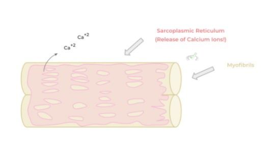
The first main commonalities between all these different types of muscle is the presence of a sarcoplasmic reticulum within the muscle fibers. This organelles is the muscle cell equivalent of the endoplasmic reticulum but holds a much more specific function in regards to muscle contraction
This organelle houses and releases an abundant amount of Ca⁺² ions which are crucial for actin-myosin cross bridge cycling which is the subcellular process that allows for muscle contraction as we cover in a more detailed article!
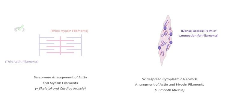
In addition, another commonality between the different types of muscle tissue is the presence of both thin, actin and thick, myosin filaments! Again, as we cover in another article, these actin and myosin protein filaments are crucial in the cross bridge cycling process resulting in muscle contraction!
However, there are differences as to how these actin and myosin filaments are arranged among the 3 types of muscle tissue. We’ll give you a quick preview but notice how skeletal and cardiac muscle have their actin and myosin filaments arranged in sarcomeres while smooth muscle has their filaments not arranged in a sarcomeric pattern but are spread out as a network throughout the cytosol.
B. Differences Between Skeletal, Cardiac, and Smooth Muscle
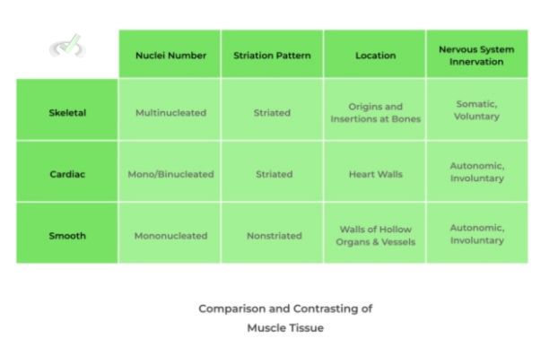
When talking about the differences among the 3 types of muscle, we’ll primarily look at contrasting the muscles in regards to the number of nuclei they have, their striation pattern, their location within the body, and their nervous system innervation!
I. Skeletal Muscle
As their name implies, skeletal muscle is associated with the skeletal system through various origins and insertions at the bones — these muscles then contract to allow for the movement of bones and body!
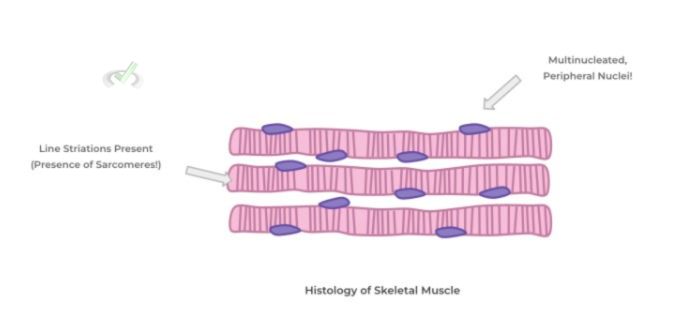
Microscopically, the skeletal muscle tissue is distinctively characterized by having multiple nuclei located on the periphery of the cells while also containing line striations within the cytoplasm — this striated pattern is due to the presence of the sarcomeric arrangement of actin and myosin filaments!
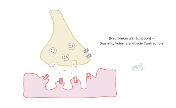
Additionally, the skeletal muscle is unique in that it’s the only type of muscle that’s under voluntary, somatic control by the nervous system.
We’ll have a whole detailed article about the processes concerning the physiological cellular and subcellular interactions that govern skeletal muscle contraction but for now, know that this takes place in a specialized synapse called the neuromuscular junction!
II. Cardiac Muscle
The name of this muscle already gives away its location as these types of muscle cells (called cardiomyocytes) reside in the heart, specifically the myocardial layer of the heart which is the contractile portion!
Histologically, the cardiac muscle is similar to skeletal muscle as they also have line striations; however, they tend to be uni or binucleated with the nucleus having a central position. Also, as shown below cardiomyocytes are connected by special gap junctions called intercalated discs which are unique to cardiac muscle tissue!
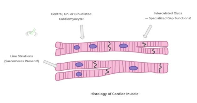
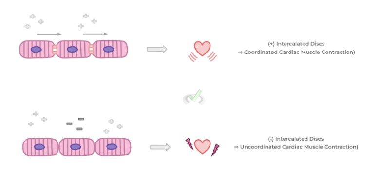
Intercalated discs are such an important feature for cardiac cardiac muscle tissue regarding the blood pumping function of the heart. The intercalated discs ensure that there’s a coordinated cardiac muscle contraction by allowing depolarization to flow from one adjacent cell to another!
If depolarization of the cardiomyocytes were to occur sporadically, then the heart chambers (such as the ventricles) wouldn’t be able to have coordinated cardiac muscle contraction resulting in an ineffective pumping of blood!
Additionally, unlike skeletal muscle, cardiac muscle is under autonomic, involuntary control! Thus, parameters like heart rate and contractility can be controlled via sympathetic and parasympathetic nerve fibers!
III. Smooth Muscle
Out of all the muscle tissues, smooth muscle is probably the most unique in terms of structure and function. For one, smooth muscle is spread throughout the body particularly residing in the GI and respiratory tract as well as blood vessels.
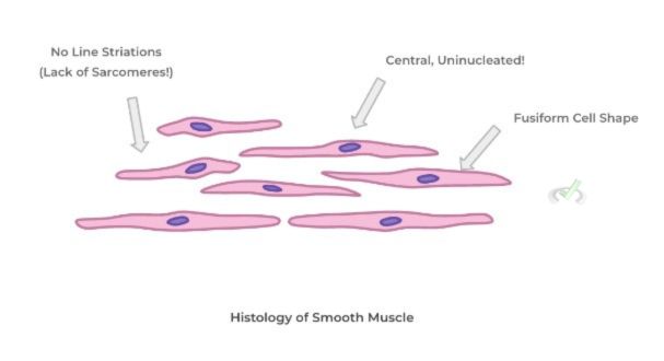
Their contraction in these various parts of the body allows for processes such as peristalsis, bronchoconstriction/dilation and vasoconstriction/dilation! Microscopically, smooth muscle cells have a single nucleus located within the cell center while also possessing a fusiform shape which kinda looks like the cell tapers towards the side ends.
These cells are also unique as they DO NOT have line striations due to their lack of sarcomeres! Recall, as we mentioned earlier, the actin and myosin filaments of smooth muscles are arranged in a network pattern throughout the cytosol as opposed to sarcomeres!
Finally, similar to çardiomyocytes, smooth muscle is also under involuntary, autonomic control of the nervous system — this makes sense in regards to their location throughout the human body as described above.
Take a look at the chart below to get a quick table summation comparing and contrasting the types of muscle!
III. Bridge/Overlap
It may be helpful to review the different divisions of the nervous system in order to better get a grasp of why the muscles are under control of a specific portion of the nervous system! Let’s go ahead and get a quick recall of the different subdivisions of the nervous system, particularly the peripheral nervous system.
I. Subdivisions of the Nervous System
The nervous system is first broken down into the central and peripheral nervous system: the central portion consists of the brain and spinal cord while the peripheral portion consists of nerves outside of the central nervous system.
The peripheral nervous system can be further subdivided into the somatic and autonomic nervous system, with the autonomic nervous system being further subdivided into the sympathetic and parasympathetic systems.
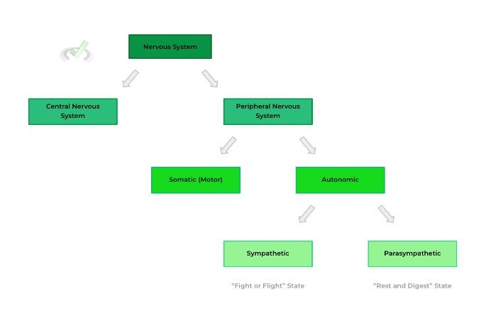
The somatic nervous system is under voluntary control which is why the skeletal muscle is stimulated by this portion of the nervous system as it takes conscious, voluntary effort in order for us to initiate skeletal muscle contraction.
Conversely, the autonomic division is under involuntary control which explains why cardiac and smooth muscle are stimulated by this division of the nervous system. It doesn’t take conscious effort for our heart to contract and pump or for our smooth muscle to contract for peristalsis to occur — there’s simply an autonomic control which allows these processes to occur without voluntary effort.
IV. Wrap Up/Key Terms
Let’s take this time to wrap up & concisely summarize what we covered above in the article!
A. Similarities Between Skeletal, Cardiac, and Smooth Muscle
All of the 3 types of muscle tissue will have a specialized organelle called the sarcoplasmic reticulum, the myofiber equivalent of an endoplasmic reticulum — this allows for the storage and release of calcium ions which is important in regards to the initiation of actin-myosin cross bridge cycling!
In addition, all the muscle tissue will have the presence of thin, actin and thick myosin protein filaments which are the proteins that interact in cross bridge cycling to allow for muscle contraction!
B. Differences Between Skeletal, Cardiac, and Smooth Muscle
When discussing the differences between skeletal, cardiac, and smooth muscle, they’re often contrasted via the amount of nuclei present, the striation pattern, their location in the body, and the type of nervous system innervation.
I. Skeletal Muscle
As the name implies, this type of muscle is associated with the bones of the skeletal system via origins and insertions. These muscles then contract via somatic, voluntary nervous stimulation which allows movement of the bone and body!
Microscopically, skeletal muscle is characterized by being peripherally multinucleated and having line striations which indicates the presence of sarcomeres.
II. Cardiac Muscle
This muscle is found in the heart, specifically the myocardial layer which allows for contraction and the pumping of blood, which is under autonomic, involuntary control!
Histologically, cardiac muscle is also striated but usually is centrally uni/binucleated. Additionally, cardiomyocytes have a specialized gap junction unique to them called intercalated discs.
The presence of the intercalated discs is crucial in order to allow for coordinated cardiac muscle contraction which allows for the efficient pumping of blood!
III. Smooth Muscle
Found throughout the body from blood vessels, GI and respiratory tract, smooth muscle allows for various autonomic physiological processes to occur such as peristalsis and vasoconstriction/dilation via their contraction!
Microscopically, smooth muscle has a single, centrally located nucleus with the cell adopting a fusiform, spindle shaped which tapers towards the side ends. Additionally, these muscle cells LACK striations as their actin and myosin filaments aren’t arranged in sarcomeres!
V. Practice
Take a look at these practice questions to see and solidify your understanding!
Sample Practice Question 1
A muscle tissue biopsy is being examined by a pathologist in order to determine the possibility of neoplasm, but there is no labeling as to what type of muscle tissue is present. Which of the following features could the pathologist use to help differentiate if the biopsy is from skeletal or cardiac muscle tissue?
I. Nuclei Number
II. Presence of Intercalated Discs
III. Striation Pattern
A. I only
B. III only
C. I and II
D. I and III
Ans. C
The nuclei number is a feature which differs between cardiac and skeletal muscle as the former is uni/binucleated while the latter tends to be multinucleated. The presence of intercalated discs is a cardiac tissue feature unique to cardiomyocytes. Finally, the striation pattern cannot be utilized to differentiate between the 2 because both have line striations indicative of sarcomeres.
Sample Practice Question 2
Albuterol is a type of medication often given to individuals who suffer from asthma, a chronic condition characterized by prolonged narrowing of the bronchial airways. Which of the following is most likely the mechanism of action of albuterol?
A. Smooth Muscle Agonist (Increase Contraction)
B. Smooth Muscle Antagonist (Decrease Contraction)
C. Skeletal Muscle Agonists (Increase Contraction)
D. Skeletal Muscle Antagonist (Decrease Contraction)
Ans. B
Recall that smooth muscle is muscle tissue that is present throughout the body but is most prominently associated with blood vessels, the GI and respiratory tract!
Because there is narrowing of the bronchial airways, this most likely means there is active contraction of the bronchial smooth muscle — as a result a bronchodilator such as albuterol can antagonize contraction of the bronchial smooth muscle and help it relax which should reduce the narrowing of the bronchial airway.





 To help you achieve your goal MCAT score, we take turns hosting these


 reviews on TrustPilot