It is crazy to think about, but everything is made up of this small unit called a cell. That means even you and I are composed of cells, over 75 trillion cells, to be exact!
Cells refer to a mass of cytoplasm that is bound externally by a cell membrane. Usually microscopic in size, can have one or more nuclei and other organelles that carry out a variety of tasks.
This guide will introduce you to the fascinating world of cells, along with some key terms and definitions you should remember as you prepare for the MCAT.
Let’s get started!
Cells on the MCAT: What Do You Need to Know?
Cells are covered in the Biology section of the MCAT.
Introductory biology accounts for 65% of the Biological and Biochemical Foundations of Living Systems section (Bio/Biochem) and 5% of the Chemical and Physical Foundations of Biological Systems section (Chem/Phys).
It’s hard to predict the exact number of questions about cells that will appear on the MCAT. However, you can expect it to appear in both the Bio/Biochem and Chem/Phys sections.
Important Sub-Topics – Cells
We cannot stress enough the importance of understanding cells during MCAT prep since this is closely related to biochemistry, and many other important MCAT topics!
Cells can be complicated, so let’s break them down into important sub-topics you can concentrate on.
1. Tissues Formed From Eukaryotic Cells
Tissues are defined as a group of structurally-related cells that come together to perform a function. Four types of eukaryotic cell tissues exist, but here are the most important ones:
Tissue Type | Description |
|---|---|
Epithelial | Composed of epithelial cells. Main function is to act as a protective layer. |
Connective | Mostly consists of ground substance, fibers, and cells, providing support for the body. |
Full Study Notes : Tissues Formed From Eukaryotic Cells on the MCAT
For more in-depth content review on tissues formed from eukaryotic cells, check out these detailed lesson notes created by top MCAT scorers.
2. Mitosis: The Cell Cycle
Our cell cycle is an organized process containing differentiating phases that are regulated via checkpoints! The cell cycle is divided into G1, S, G2, and M phases with a quiescent G0 state while being regulated for possible errors by many checkpoints.
A. G1 Phase
In this phase, the cell grows in both size and number of organelles to eventually reach an even distribution of each into the two daughter cells.

B. S (synthesis) Phase
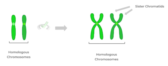
This phase involves the synthesis and replication of DNA. This replication of DNA contains 2 genetically identical sister chromatids held together by a centromere.
C. G2 Phase
In this phase, the cell continues to grow and synthesizes many mitotic proteins. At this stage if any DNA damage is detected, entrance to M phase is halted until endonucleases and exonucleases repair the damage.

D. M (mitotic) Phase
This phase results in the formation of two genetically identical daughter cells from one parent cell. Has four phases – prophase, metaphase, anaphase, and telophase.

E. G0 (Growth arrest) Phase
Sometimes, cells enter this phase when not actively pursuing to divide, such as neuronal cells.
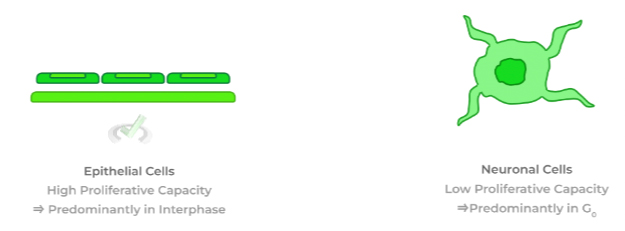
There are also two checkpoints – G1/S and G2/M checkpoints to regulate the cell cycle pathway, ensuring progression through the cell cycle is functioning properly.
Full Study Notes : Mitosis: The Cell Cycle on the MCAT
For more in-depth content review on the mitosis cell cycle, check out these detailed lesson notes created by top MCAT scorers.
3. Mitosis: Structures and Processes
There are four phases of mitosis – prophase, metaphase, anaphase and telophase.
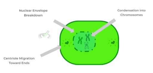
Phase 1: Prophase
This involves the degradation of the nuclear envelope.
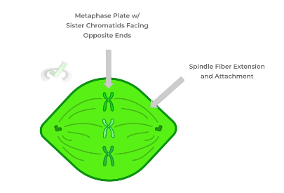
Phase 2: Metaphase
In metaphase, the condense chromosomes begin to migrate towards the midline of the cell.
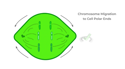
Phase 3: Anaphase
Separation of sister chromatids via retraction of spindle fibers.
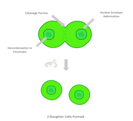
Phase 4: Telophase
Nuclear envelope reforms and chromosomes decondense back to chromatin.
Full Study Notes : Mitosis: Structures and Processes on the MCAT
For more in-depth content review on the mitosis structures and processes, check out these detailed lesson notes created by top MCAT scorers.
4. Meiosis: Significance and General Overview
Meiosis is the basis for how we reproduce as species! Meiosis results in gametes or sex cells that fuse together during fertilization, generating a genetically unique zygote.
Human gametes are diploid, meaning we have two sets of chromosomes. In total, we have 23 pairs of homologous chromosomes or 46 chromosomes.
Meiosis differs from mitosis since meisosis produces 4 gamete sex cells each with half the amount of genetic information of the parent cell. Whereas mitosis produces two genetically identical daughter cells. Meiosis also occurs in two phases, whereas mitosis occurs in only one.
(Full Study Notes : Meiosis: Significance and General Overview
For more in-depth content review on the significance and general overview, check out these detailed lesson notes created by top MCAT scorers.
5. Meiosis: Processes
As mentioned previously, meiosis has two phases – Meiosis I and Meiosis II.
Let's begin with meiosis I: segregation of homologous chromosomes
Phase | Description |
|---|---|
Prophase I | Degradation of the nuclear envelope, chromosome condensation, and genetic recombination occur due to crossing over, which increases genetic diversity. |
Metaphase I | The different combinations of chromosome alignment on the metaphase plate can increase genetic diversity. |
Anaphase I | Homologous chromosomes are being separated (different from mitosis!) |
Telophase I | Two haploid daughter cells are generated, each only containing one set of chromosomes. |
Following meiosis I, is meiosis II: segregation of sister chromatids and genes
Phase | Description |
|---|---|
Prophase I | Similar to prophase I, there is the disappearance of the nuclear envelope, and chromosomes condense. |
Metaphase I | Sister chromatids align at the metaphase plate. |
Anaphase I | Sister chromatids are separated |
Telophase I | Four haploid genetically unique daughter cells are formed |
Full Study Notes : Meiosis: Processes
For more in-depth content review on the meiosis: processes, check out these detailed lesson notes created by top MCAT scorers.
6. Prokaryotic cells: General Structural and Physiological Characteristics
Prokaryotic cells are somewhat similar to eukaryotic cells but have a list of characteristics unique to them:
- Lack of nucleus and mitotic features such as spindle fibers and centrioles since prokaryotic cells carry out binary fission
- Lack of membrane-bound organelles
- Presence of a bacterial cell wall and composition
- Presence of flagella allow for bacterial locomotion
- Have symbiotic relationships between the bacteria and the host (mutualism, commensalism, and parasitism)
Full Study Notes : Prokaryotic Cells: General Structural and Physiological Characteristics on the MCAT
For more in-depth content review on the prokaryotic cells’ general structure and physiological characteristics, check out these detailed lesson notes created by top MCAT scorers.
7. Prokaryotic Cells: Cell Theory and Major Classifications
Cell theory was discovered by Robert Hooke and has 4 main tenets:
- Cells are the basic structural units of life.
- All living organisms are composed of cells.
- Cells can only arise from pre-existing cells and do so independently.
- DNA is used to transmit the necessary genetic codes, which contain the information needed for cell growth and survival.
Cells can also be classified in many different ways, such as cell domains, anaerobic vs. aerobic metabolism, and shape.
Full Study Notes : Prokaryotic Cells: Cell Theory and Major Classifications on the MCAT
For more in-depth content review on the cell theory and major classifications of prokaryotic cells, check out these detailed lesson notes created by top MCAT scorers.
8. Prokaryotic Cells: Cell Growth
In order for cell growth to occur, we must undergo mitosis. However, in prokaryotes, mitosis does not occur. Instead, there is a similar cellular mechanism called binary fission. This process allows a bacterial cell to divide into 2 genetically identical daughter cells.
Due to the simplicity of binary fission, bacterial growth can actually be modeled as an exponential function, which has four phases:
- Lag Phase: Represents when bacteria are not yet dividing and acclimating to the new environment.
- Exponential (Log) Phase: Represents the stage of rapid, exponential bacterial cell growth.
- Stationary Phase: Represents the slowing of reproduction due to the depletion of nutrients and/or resources.
- Death (Decay) Phase: Represents bacterial death due to the environment no longer being able to support cell growth.
Full Study Notes : Prokaryotic Cells: Cell Growth on the MCAT
For more in-depth content review on the cell growth of prokaryotic cells, check out these detailed lesson notes created by top MCAT scorers.
9. Prokaryotic Cells: Genetic Exchange and Recombination
Bacterium can have extragenomic DNA plasmids outside of their typical circular DNA chromosome.

These extragenomic DNA plasmids have genes that code for specialized functions such as antibiotic resistance!
Gene transfer can occur both horizontally and vertically. Meaning the transfer is occurring within the same generation or different generations, respectively.
For an example, we’ll look at the steps for a horizontal gene transfer (within the same generation of bacterium):
Step 1 - Transformation:
This is described as the uptake of extragenomic DNA from the environment.

Step 2 - Conjugation:
This is a gene transfer that occurs through the use of a sex pilus structure.

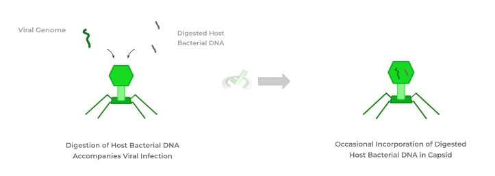
Step 3 - Transduction:
The use of a bacteriophage to insert foreign bacterial DNA into a bacterium.
Full Study Notes : Prokaryotic Cells: Genetic Exchange and Recombination on the MCAT
For more in-depth content review on the genetic exchange and recombination of prokaryotic cells, check out these detailed lesson notes created by top MCAT scorers.
10. Prokaryotic Cells: Transposons
Transposons are DNA sequences that can switch/translocate between different positions in the genome.
The main structure of a transposon is a transposase gene sandwiched between two inverted terminal repeat (ITR) ends.
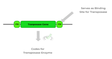
Transposase utilizes the ITRs as binding recognition sites to cut out the transposon. Afterward, transposase cuts at another DNA sequence site which allows for the insertion of the transposon at the new location.

Full Study Notes : Prokaryotic Cells: Transposons on the MCAT
For more in-depth content review on transposons, check out these detailed lesson notes created by top MCAT scorers.
11. Viruses: General Structural Characteristics
Viruses have two main components – a capsid protein shell and a nucleic acid genome. Additionally, depending on the presence of a lipid membrane enclosing the virus, the virus can be determined as enveloped or nonenveloped.
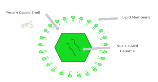
Viruses break the 4th tenet of cell theory, as they can have both DNA and RNA genomes that can be either single or double-stranded.
Single-stranded RNA genomes can either be (+) or (-) sense. If the RNA genome can be translated immediately, it is a (+)-sense genome. A (-)-sense genome must first utilize an RNA-dependent RNA polymerase to transcribe a complementary (+)-sense strand that can undergo translation.
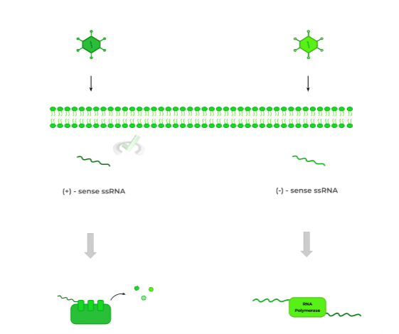
Another key characteristic of viruses is their lack of nuclei and organelles. Additionally, viruses are extremely small, much smaller than prokaryotes and eukaryotes.
Full Study Notes : Viruses: General Structural Characteristics on the MCAT
For more in-depth content review on the general characteristics of viruses, check out these detailed lesson notes created by top MCAT scorers.
12. Steps of the Viral Cycle
The viral cycle occurs in a series of steps. Both the phage and animal viruses share general steps:
A. Entry
Attachment occurs via protein-receptor binding for animal viruses and through tail fiber anchoring for general phages. Animal viruses infiltrate the cell through either membrane fusion, endocytosis, or genome insertion.
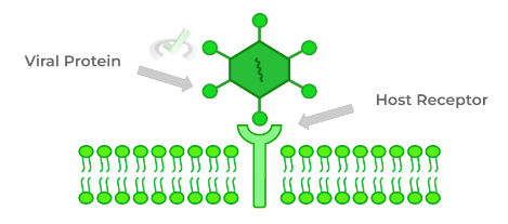
B. Transcription, translation, and genome replication
Transcription and translation come first to create necessary proteins for viral infection such as capsid proteins and restriction enzymes. Following this, genome replication occurs.

C. Assembly: self-replicating biological units
The replicated viral components then self-assemble to form virions.

D. Release
As all the virions come together, they are then released via cell lysis, killing the cell.
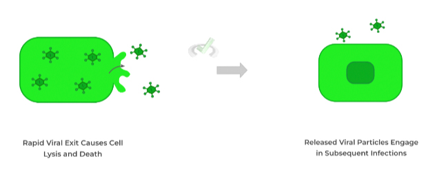
Full Study Notes : Viruses: Steps of the Viral Life Cycle on the MCAT
For more in-depth content review on the steps of the viral cycle, check out these detailed lesson notes created by top MCAT scorers.
13. Subviral Particles: Prions and Viroids
Prions and viroids are a relatively new discovery and, therefore, a somewhat low-yield topic on the MCAT:
Subviral Particle | Description |
|---|---|
Prions | Misfolded infectious proteins give rise to some neurodegenerative diseases. |
Viroids | Infectious circular RNA molecules that cause RNA silencing in plant cells. |
Full Study Notes : Viruses: Subviral Particles Prions and Viroids on the MCAT
For more in-depth content review on subviral particles, check out these detailed lesson notes created by top MCAT scorers.
Key Terms and Definitions – Cells
Here are some of the more important key terms and definitions to remember for this general guide to cells!
Term | Definition |
|---|---|
Centromeres | Links a pair of sister chromatids together during cell division. |
Spindle Fibers | A protein structure that divides the genetic material in a cell. |
Homologous Chromosomes | Two chromosomes in a pair, one inherited from the mother and one from the father. |
Bacteriophage | Viruses that infect bacterial cells. They have tail sheath and tail fiber structures. The tail fibers anchor the phage to the bacteria while the tail sheath injects the viral genome into the bacterium, initiating infection. |
Binary Fission | Cell divides asexually to produce two daughter cells. |
Genetic Recombination | Exchange of genetic information leading to offspring with traits that differ from those found in the parents. |
Additional FAQs – Cells on the MCAT
Is Cell Biology On The MCAT?
What Are The 12 Organelles In A Cell?
Additional Reading Links – Study Notes for Cells on the MCAT
For more in-depth content review about cells on the MCAT, check out these detailed lesson notes created by top MCAT scorers!
Additional Reading: MCAT Biology Topics:
- Cardiovascular Systems on the MCAT
- Digestive Systems on the MCAT
- Embryogenesis and Development on the MCAT
- Endocrine Systems on the MCAT
- Excretory Systems on the MCAT
- Genetics and Evolutions on the MCAT
- Immune Systems on the MCAT
- Nervous Systems on the MCAT
- Musculoskeletal Systems on the MCAT
- Reproduction on the MCAT
- Respiratory Systems on the MCAT


 To help you achieve your goal MCAT score, we take turns hosting these


 reviews on TrustPilot