Studying the brain helps us understand how it works and what happens when things go wrong. Brain imaging techniques give us a window into this complex organ.
These methods allow scientists and doctors to see the brain's structure, function, and chemical composition. Let's explore the various methods used for brain imaging and studies.
I. Structural Imaging
Structural imaging methods provide detailed pictures of the brain's anatomy. They help identify physical abnormalities, such as tumors or brain injuries.
A. Magnetic Resonance Imaging (MRI)
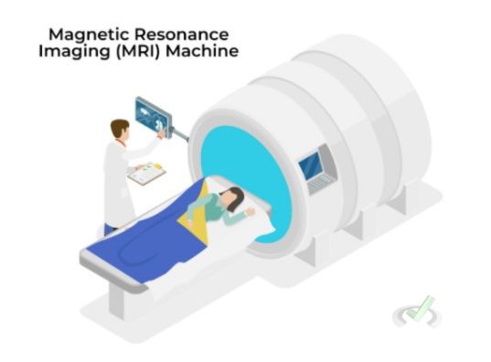
MRI uses powerful magnets and radio waves to create detailed brain images.
How MRI Works:
- The brain contains water molecules (H₂O).
- The hydrogen nuclei (protons) in water align with the magnetic field. This alignment depends on the Larmor equation: ω = γB, where ω is the angular frequency, γ is the gyromagnetic ratio, and B is the magnetic field strength.
- Radio waves disrupt this alignment, and as the protons return to their original state, they emit signals.
- These signals are captured and converted into images by a computer.
Uses:
- Detecting brain tumors and multiple sclerosis lesions.
- Identifying brain injuries and developmental disorders.
- Mapping brain structure and abnormalities.
B. Computed Tomography (CT)
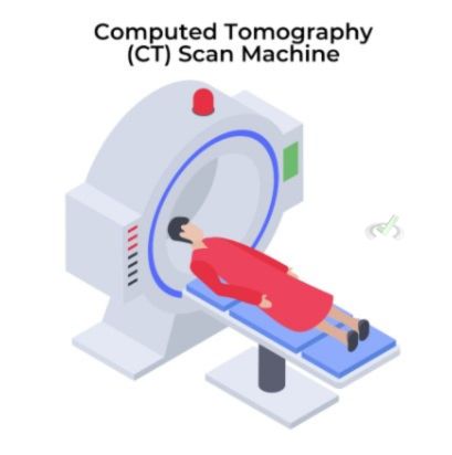
CT scans use X-rays to create detailed cross-sectional images of the brain.
How CT Works:
- X-rays pass through the brain at different angles.
- Different tissues absorb X-rays differently, creating contrast in the images.
- A computer compiles these images to create a detailed cross-sectional view.
- This method is useful for quickly diagnosing brain injuries.
Uses:
- Detecting bleeding in the brain (hemorrhages).
- Identifying skull fractures and brain injuries.
- Assessing structural abnormalities and tumors.
C. Diffusion Tensor Imaging (DTI)
DTI is a type of MRI that maps the diffusion of water molecules in brain tissue, highlighting neural pathways.
How DTI Works:
- Measures the movement of water molecules in the brain.
- Creates images of white matter tracts, showing how different brain regions are connected.
- Helps understand brain connectivity and structural integrity.
Uses:
- Studying brain development and aging.
- Investigating brain injuries and neurodegenerative diseases.
- Mapping neural pathways for surgical planning.
II. Functional Imaging
Functional imaging techniques show brain activity. These methods help us understand how different parts of the brain work during various activities.
A. Functional MRI (fMRI)
fMRI measures brain activity by detecting changes in blood flow.
How fMRI Works:
- Active brain areas require more oxygen, which increases blood flow to these regions.
- The Blood Oxygen Level Dependent (BOLD) signal changes the magnetic properties of blood.
- fMRI detects these changes, indicating brain activity.
Uses:
- Studying brain functions during tasks like reading or problem-solving.
- Understanding brain disorders such as depression and schizophrenia.
- Mapping brain activity for surgery planning, particularly in regions involved in critical functions like speech.
B. Positron Emission Tomography (PET)
PET scans use radioactive tracers to visualize brain activity.
How PET Works:
- A radioactive tracer, such as Fluorodeoxyglucose (FDG), is injected into the bloodstream.
- The tracer accumulates in active brain areas, which use more glucose.
- The PET scanner detects the emitted radiation, showing brain activity.
Uses:
- Studying brain metabolism and activity.
- Detecting Alzheimer's disease by identifying amyloid plaques.
- Mapping brain functions and diagnosing conditions like epilepsy.
C. Electroencephalography (EEG)
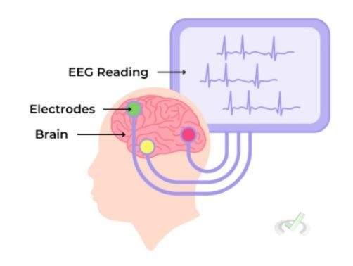
EEG records electrical activity in the brain using electrodes placed on the scalp.
How EEG Works:
- Electrodes detect electrical signals produced by brain cells (neurons).
- These signals are amplified and recorded by a computer.
- EEG provides real-time data on brain activity.
Uses:
- Diagnosing epilepsy and other seizure disorders.
- Studying sleep disorders.
- Monitoring brain activity during surgery.
D. Near-Infrared Spectroscopy (NIRS)
NIRS measures brain activity by detecting changes in blood oxygen levels using near-infrared light.
How NIRS Works:
- Near-infrared light is emitted into the scalp.
- The light penetrates the skull and is absorbed by blood.
- Light absorption changes indicate blood oxygen levels, reflecting brain activity.
Uses:
- Studying brain function in infants and young children.
- Researching brain activity in naturalistic settings.
- Monitoring brain function in critical care.
E. Magnetoencephalography (MEG)
MEG measures the magnetic fields that are produced by brain activity.
How MEG Works:
- Sensors detect magnetic fields generated by neuronal activity.
- Provides real-time data on brain activity with high temporal resolution.
- Non-invasive and silent.
Uses:
- Studying brain functions, such as sensory processing and motor control.
- Localizing brain regions involved in specific tasks.
- Pre-surgical planning for epilepsy treatment.
III. Molecular Imaging
Molecular imaging provides insights into the brain's chemical processes. This method is essential for studying neurotransmitters and brain chemistry.
A. Single Photon Emission Computed Tomography (SPECT)
SPECT uses gamma rays to create detailed images of brain activity.
How SPECT Works:
- A radioactive tracer gets injected into the bloodstream.
- The tracer emits gamma rays while it decays.
- SPECT captures these gamma rays to create images of brain activity.
Uses:
- Studying brain blood flow and metabolism.
- Diagnosing epilepsy by identifying seizure foci.
- Assessing brain function after a stroke or head injury.
B. Magnetic Resonance Spectroscopy (MRS)
MRS measures chemical levels in the brain.
How MRS Works:
- Uses similar technology to MRI but focuses on chemical composition.
- Measures levels of neurotransmitters and other chemicals, such as N-acetyl aspartate (a marker for neuronal health) and choline (a marker for cell membrane turnover).
Uses:
- Studying brain metabolism and chemical imbalances.
- Investigating brain tumors by identifying their chemical profiles.
- Understanding neurological disorders like multiple sclerosis.
IV. Bridging Concepts
Brain imaging techniques help diagnose diseases and advance our understanding of the brain. These methods contribute to research in neuroscience, psychology, and medicine.
A. Integrating Structural and Functional Imaging
Combining different imaging techniques provides a comprehensive view of the brain. For example:
- MRI and fMRI together show both structure and function.
- CT and PET scans offer insights into brain anatomy and activity.
B. Advances in Brain Imaging
New technologies continue to improve brain imaging:
- High-resolution MRI for detailed brain maps, improving the diagnosis of small lesions.
- Real-time fMRI for immediate feedback on brain activity, used in neurofeedback therapies.
- Hybrid PET/MRI scanners enhance diagnostic accuracy for combined metabolic and structural information.
C. Understanding Brain Disorders
Brain imaging helps us understand and treat various brain disorders:
- Alzheimer's disease: PET scans detect amyloid plaques and monitor disease progression.
- Depression: fMRI studies brain activity changes, aiding in identifying specific brain regions involved.
- Epilepsy: SPECT pinpoints seizure origins, assisting in surgical planning.
D. Connecting Brain Imaging to Organic Chemistry
Brain imaging and organic chemistry intersect in understanding the chemical basis of brain function and disease. For example:
- Neurotransmitter Chemistry: PET and MRS can study neurotransmitters like dopamine and serotonin, which are organic molecules.
- Chemical Kinetics: Understanding how radioactive tracers decay and their interaction with brain tissues involves principles of chemical kinetics.
V. Wrap-Up and Key Terms
Brain imaging techniques provide critical insights into the brain's structure, function, and chemistry. These methods are vital for diagnosing brain disorders and advancing our understanding of how the brain works.
Key Terms
- MRI (Magnetic Resonance Imaging): Uses magnets and radio waves to create brain images.
- CT (Computed Tomography): Uses X-rays for cross-sectional brain images.
- fMRI (Functional MRI): Measures brain activity via blood flow changes.
- PET (Positron Emission Tomography): Uses radioactive tracers to show brain activity.
- SPECT (Single Photon Emission Computed Tomography): Uses gamma rays to visualize brain activity.
- MRS (Magnetic Resonance Spectroscopy): Measures chemical levels in the brain.
VI. Practice Questions
Sample Practice Question 1
A patient shows signs of early-stage Alzheimer's disease. Which imaging technique is likely to be used to detect amyloid plaques in the brain?
A. MRI
B. CT
C. PET
D. SPECT
Ans. C
PET scans use radioactive tracers to visualize brain activity and can detect amyloid plaques indicative of Alzheimer's disease.
Sample Practice Question 2
During a functional study, researchers want to observe changes in blood flow to specific brain regions as a participant solves math problems. Which imaging technique should they use?
A. MRI
B. fMRI
C. CT
D. MRS
Ans. B
fMRI measures brain activity by detecting changes in blood flow, making it suitable for observing brain functions during tasks.




 To help you achieve your goal MCAT score, we take turns hosting these




 reviews on TrustPilot