I. What are the Steps in Digestion?
When you chow down on a delicious meal of ramen, Korean barbeque, a nice salad, or any one of your favorite meals, the process of digestion begins immediately!
Without you even knowing or making a conscious effort, your body engages in the necessary processes of breaking down food and absorbing all the nutrients needed to keep your organs, tissues, and cells functioning properly—let’s dive in and look into the process of digestion!
II. The Process of Digestion
In general, and for the purposes of the MCAT, we’ll divide digestion into three main categories and steps: mechanical digestion, chemical digestion, and absorption.
A. Mechanical Digestion
This step of digestion can also be best thought of as the “increasing surface area” portion! Essentially, the body is taking in the ingested food and trying to use mechanical, shearing forces to break down the food particles and increase their surface area for digestion.
The 2 most common examples of this mechanical process are mastication (i.e. chewing food) and peristalsis — while we’re all familiar with the process of chewing, peristalsis is a unique concept that’s not as intuitive.
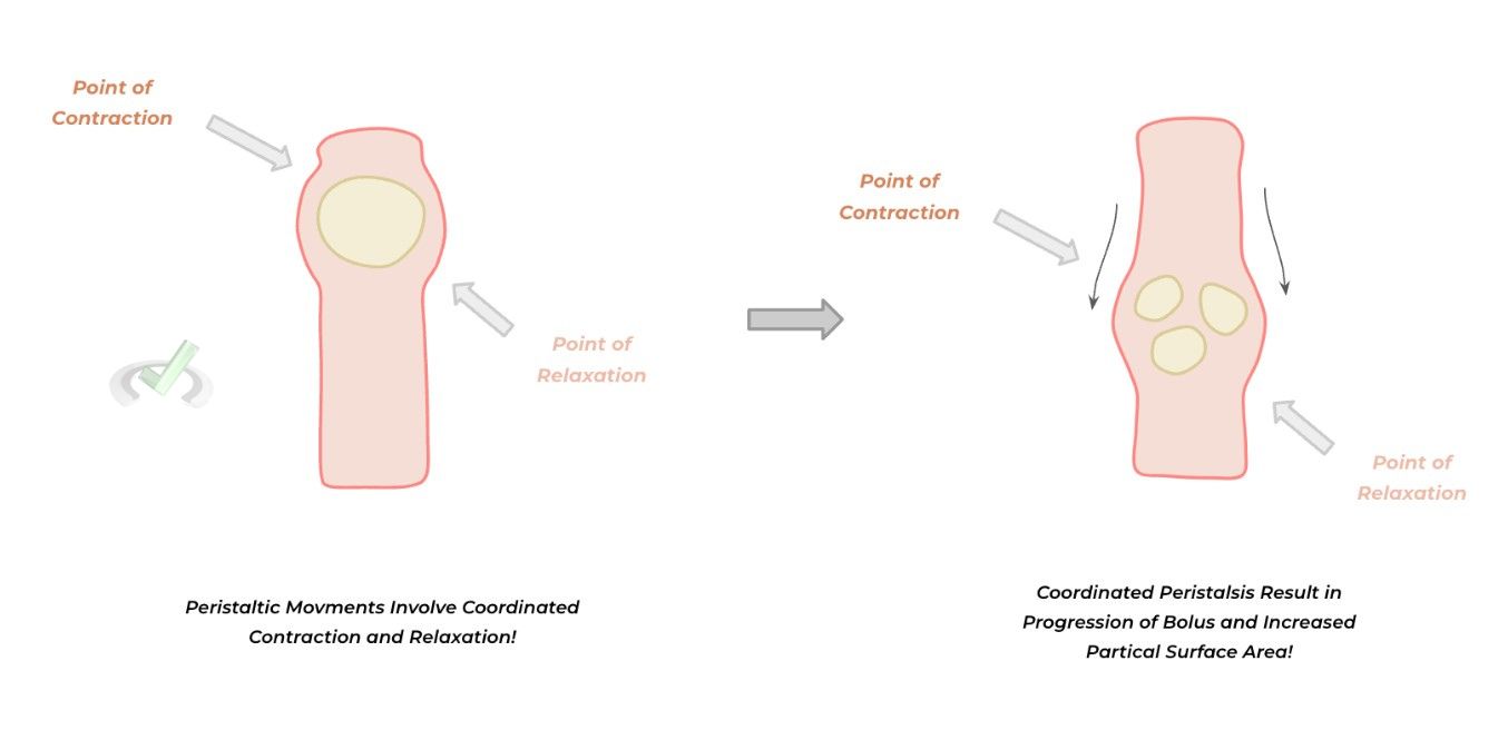
This process refers to when the smooth muscle surrounding the GI tract continues to contract and relax in a rhythmic fashion, which not only progresses the food forward but also mechanically grinds and shears the food bolus to increase the surface area.
I. Bile Salts
While bile salts aren’t technically a source of mechanical digestion, they do indeed have an important role in increasing the surface area for digestion, particularly incoming fat droplets!
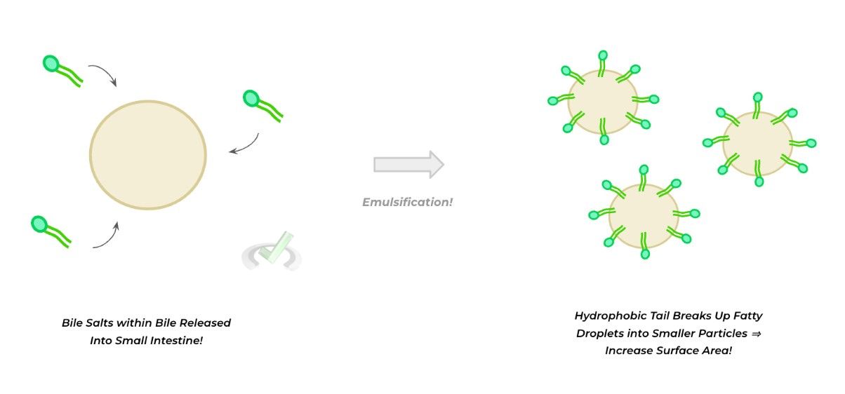
Contained within bile are what are called bile salts, which are amphipathic molecules containing a hydrophilic head (usu. glycine) and a hydrophobic tail composed of a cholesterol derivative.

When fat droplets reach the small intestine, the bile salts—via their hydrophobic tail—disrupt the integrity of the fatty droplets by encompassing them and making them smaller fat droplets in a process called emulsification, which increases their surface area.
B. Chemical Digestion
As the name implies, this step of digestion also involves the breakdown of the incoming food particles; however, in this particular form, we’re looking at how various enzymes within the GI tract act upon substrates to metabolize them.
In this step, we’ll break down chemical digestion based on the enzyme as well as what product is being broken down: 1) proteolytic degradation, 2) fat digestion, and 3) carbohydrate metabolism.
I. Proteolytic Degradation
There are a variety of proteolytic enzymes within the gastrointestinal tract that act upon incoming proteins to break them down into smaller and easier-to-be-absorbed amino acids/oligopeptides.
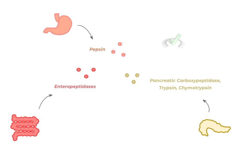
These include pepsin (secreted within the stomach), carboxypeptidases, chymotrypsin and trypsin (secreted by the pancreas), and enteropeptidases (brush border proteolytic enzymes in the small intestine).
It’s also important to note that these proteolytic enzymes are secreted as inactive precursors called zymogens named as follows:
- Pepsinogen ⇒ Pepsin
- Chymotrypsinogen ⇒ Chymotrypsin
- Trypsinogen ⇒ Trypsin
- Procarboxypeptidase ⇒ Carboxypeptidase
- Proenteropeptidases ⇒ Enteropeptidase
Generally, these precursor enzymes are configured in a way where their active site is inaccessible.
As such, it’s only during active digestion that the enzymes can be altered and activated (i.e. proteolytic cleavage from another enzyme, change in pH) that the zymogens can be converted into their active form and actively engage in digestion!
II. Fat Digestion
The primary enzyme for digesting fats is pancreatic lipase, which is also stored within the pancreatic fluid.
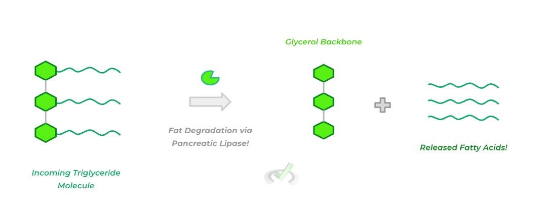
After the released bile salts increase the surface area for the incoming fat droplets, pancreatic lipase primarily targets incoming triglycerides and hydrolyzes them in order to release the esterified fatty acids and glycerol!
III. Carbohydrate Metabolism
Finally, there are various enzymes important for the metabolism of both simple and complex carbohydrates!
In addition to enteropeptidases, various disaccharidases break down incoming disaccharides! These include maltase (breaking down maltose), sucrase (breaking down sucrose), and lactase (breaking down lactose).
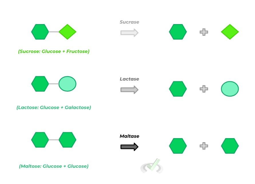
Finally, the pancreatic fluid also houses pancreatic amylase which is primarily responsible for breaking down complex carbohydrates as seen in fibers and whole grains. There is also salivary amylase released by the salivary glands which also aids in this process.
C. Absorption
Finally, after all the incoming food particles are broken down, the various epithelial cells of the gastrointestinal lining absorb the broken-down food, which is then transported in the bloodstream and distributed throughout the various tissues of the body.
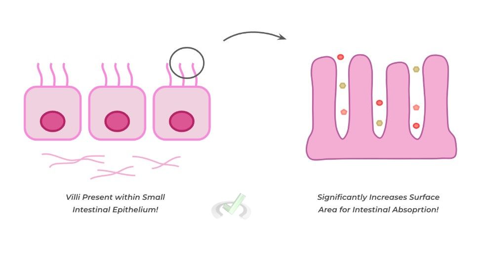
It’s important to note that much of the absorption of water, electrolytes, and nutrients occurs in the small intestine due to the presence of villi. These fingerlike projections extend from the GI epithelium and greatly increase the surface area for absorption!
While there are a multitude of transport proteins involved in gut absorption, the MCAT definitely won’t hold you accountable for memorizing those (believe us, you’ll have plenty of time for that in medical school 
As of now, probably the 2 most high-yield to know or at least have some familiarity with are the sodium-glucose cotransporter (SLGT1) and the GLUT2 transporter.
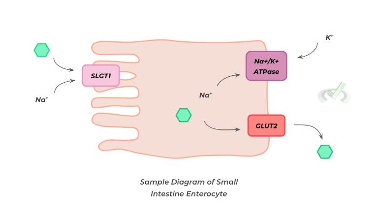
Utilizing the downstream passive diffusion of sodium, the SLGT1 (located on the apical side) can provide the energy to move glucose up its concentration gradient. The GLUT2 transporter, located on the basolateral side, transports the glucose into the bloodstream.
Finally, it’s important to note that while the large intestine has the sole function of absorbing water (and water-soluble vitamins), most water absorption is done in the small intestine.
III. Bridge/Overlap
While it may not seem intuitive at first sight, the emulsification of incoming fat droplets via bile salts is actually a great real-life application of the hydrophobic effect! Let’s go ahead and review these concepts real quick!
I. The Hydrophobic Effect
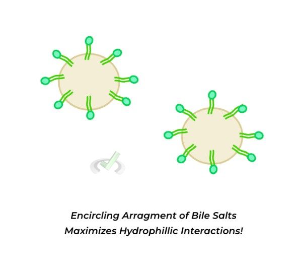
Recall that the hydrophobic effect refers to the chemical behavior where hydrophobic molecules tend to aggregate together to maximize hydrophilic interactions—this is ultimately due to the differences in polarity.
As shown below, the bile salts conform so that hydrophobic tails encircle the fat droplets, maximizing the interactions of hydrophilic molecules with each other!
IV. Wrap Up/Key Terms
Let’s take this time to wrap up & concisely summarize what we covered above in the article!
A. Mechanical Digestion
The body can utilize mechanical forces to effectively maximize the surface area for digestion. These can include mastication (chewing) or the rhythmic peristaltic movements of the gastrointestinal tract.
I. Bile Salts
In addition to the above mechanisms, bile salts stored and secreted by the gallbladder can encircle incoming fat droplets in order to increase their surface area for fatty degradation.
B. Chemical Digestion
This type of digestion involves the action of various enzymes in order to break down incoming food particles into smaller and easily digestible metabolites.
I. Proteolytic Degradation
There are various types of proteolytic enzymes that are released by the various cells of the GI tract in order to break down incoming proteins into smaller amino acids/oligopeptides.
These proteolytic enzymes are released in their inactive forms called zymogens and are activated through various mechanisms such as changes in pH or another proteolytic enzyme enacting upon it. A list of the common ones is given below:
- Pepsinogen ⇒ Pepsin
- Chymotrypsinogen ⇒ Chymotrypsin
- Trypsinogen ⇒ Trypsin
- Procarboxypeptidase ⇒ Carboxypeptidase
- Proenteropeptidases ⇒ Enteropeptidase
II. Fat Digestion
The main enzyme responsible for fat digestion is pancreatic lipase which is also contained within the pancreatic fluid.
It acts upon incoming triglycerides and hydrolyzes the ester bonds to release the glycerol backbone and the fatty acids, which can then be absorbed!
III. Carbohydrate Metabolism
Finally, various brush-border disaccharidases can break down simple disaccharides. These include maltase (breaking down maltose), sucrase (breaking down sucrose), and lactase (breaking down lactose).
In addition, salivary and pancreatic amylase (released by the salivary glands and the pancreas, respectively) aid in the breakdown of complex carbohydrates.
C. Absorption
Most of the absorption of water, electrolytes, and metabolites occurs in the small intestine. This is due to the presence of villi within the small intestine epithelium, which allows for a significant increase in surface area for absorption along the small intestinal tract.
The 2 main transport proteins are best to be familiar with for the MCAT: the SGLT-1 cotransporter and the GLUT 2 transporter.
The SLGT-1 protein (on the apical side) utilizes the downstream movement of sodium in order to power the upstream movement of glucose into the cell. The GLUT 2 transporter (on the basolateral side) transports glucose into the bloodstream to be distributed throughout the body.
V. Practice
Take a look at these practice questions to see and solidify your understanding!
Sample Practice Question 1
A new patient comes into her primary care physician complaining of repeated bouts of watery diarrhea. Upon further history taking, the physician learns that the patient’s episodes of watery diarrhea usually occur after ingesting products high in lactate such as dairy products, ice cream, etc. The physician suspects a deficiency in a crucial digestive enzyme: the suspected enzyme that’s deficient is best described as what type of enzyme?
A. Disccharidase
B. Lipase
C. Amylase
D. Protease
Ans. A
The enzyme which is most likely deficient is lactase, which recall is one of the disaccharidases whose main job is to break down lactate into glucose and galactose.
Recall as well that the disaccharidases include sucrase (which breaks down sucrose into glucose and fructose) and maltase (which breaks down maltase into 2 glucose molecules).
Sample Practice Question 2
A patient who suffers from gastroesophageal reflux disease (GERD) is prescribed proton pump inhibitors in order to decrease the acid secretion within the stomach. However, the doctor is wary that the decreased acidity of the stomach might prevent the activation of a crucial digestive enzyme. Which of the following is the name of the inactive enzyme that may predominate in the stomach if PPIs are given to the patient?
A. Chymotrypsinogen
B. Pepsinogen
C. Trypsinogen
D. Procarboxypeptidase
Ans. B
Recall that pepsinogen is the zymogen secreted in the stomach via chief cells as the inactive form of pepsin — it is primarily due to the increased acidity within the stomach (due to HCl secretion from the parietal cells) that pepsinogen is activated to its main proteolytic form as pepsin.



 To help you achieve your goal MCAT score, we take turns hosting these





 reviews on TrustPilot